
Fig. 2
- ID
- ZDB-IMAGE-200414-2
- Publication
- Ward et al., 2019 - Pharmacological restoration of visual function in a zebrafish model of von-Hippel Lindau disease
- All Figures
- Figures for Ward et al., 2019
|
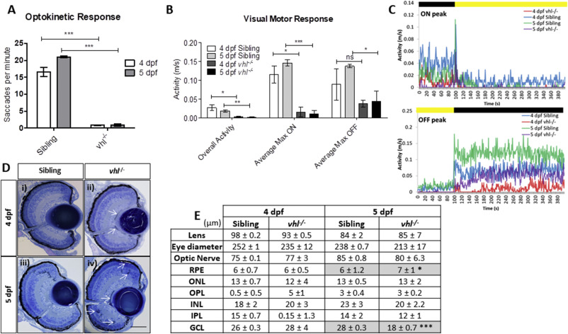
Fig. 2 vhl−/− larvae display retinal abnormalities and impaired visual function. vhl−/−larvae possess A) reduced visual capacity in response to a moving striped stimulus (OKR) n = 3, 12 technical replicates per treatment group per experiment. B) reduced movement in response to changes in light intensity (VMR) and C) VMR chromatographs illustrating average max ON/OFF peaks. n = 3, 12 technical replicates per treatment group per experiment. D) vhl−/− larvae display defects in retinal arrangement at 5 dpf. Representative light microscopy images of retinal sections from i) 4 dpf sibling control, ii) 4 dpf vhl−/− iii) 5 dpf sibling control and iv) extensive retinal damage, thought to be fluid accumulation present in 5 dpf vhl−/− larvae (white arrows). Scale bar = 50 μm. E) Morphometric analysis based on 3 independent measurements from 3 larvae per group. Values represent mean ± standard deviation of retinal layer thickness in μm. No statistically significant differences is observed between retinal layers of sibling and vhl−/− at 4 dpf. The retinal layers analysed include Retinal Pigment Epithelium (RPE), Outer Nuclear Layer (ONL), Outer Plexiform Layer (OPL), Inner Nuclear Layer (INL), Inner plexiform Layer (IPL) and Ganglion Cell Layer.
Reprinted from Developmental Biology, 457(2), Ward, R., Ali, Z., Slater, K., Reynolds, A.L., Jensen, L.D., Kennedy, B.N., Pharmacological restoration of visual function in a zebrafish model of von-Hippel Lindau disease, 226-234, Copyright (2019) with permission from Elsevier. Full text @ Dev. Biol.