
Fig 4
- ID
- ZDB-IMAGE-200412-14
- Publication
- Xie et al., 2020 - E2f5 is a versatile transcriptional activator required for spermatogenesis and multiciliated cell differentiation in zebrafish
- All Figures
- Figures for Xie et al., 2020
|
Fig 4
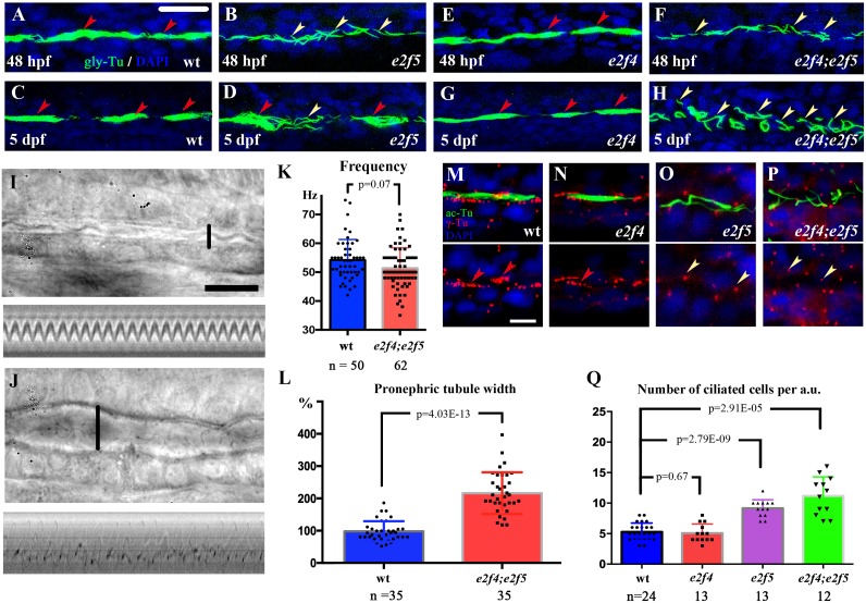
(A-H) Confocal images showing cilia in the PST region of the pronephros of wild-type and mutant larvae at different stages as indicated. Cilia were visualized with anti-glycylated tubulin antibody in green. Nuclei were labeled with DAPI in blue. Red arrowheads indicate multicilia bundles and yellow arrowheads indicate single cilia. (I-J) Still images from