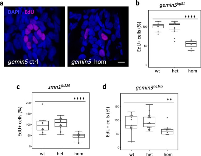
Decreased neuromast cell proliferation after hair cell ablation in gemin3, gemin5, and smn1 mutants.
a Confocal images of lateral line neuromasts in the control and gemin5hg81 mutant at 1-day post hair cell ablation. Neuromasts were stained with DAPI (blue) and proliferating cells were labeled by EdU (pink). The embryos used for the analysis were obtained from a pairwise incross of heterozygotic parents. Hair cells were ablated at 5 dpf. EdU treatment was conducted at 1-day post ablation. Scale bar, 10 µm. b Quantification of EdU-positive cells in the embryos carrying wild-type, heterozygous, or homozygous gemin5hg81 mutations. c Quantification of EdU-positive cells in the embryos carrying wild-type, heterozygous, or homozygous smn1fh229 mutations. d Quantification of EdU-positive cells in the embryos carrying wild type, heterozygous, or homozygous gemin3hg105 mutations. The graphs show mean ± s.e.m. Homozygous mutants for all three regeneration genes have a significantly reduced number of proliferating cells. **P < 0.01; ****P < 0.0001. Data for each mutation were generated using ~40 embryos born from a single pair of heterozygous parents.
This image is the copyrighted work of the attributed author or publisher, and
ZFIN has permission only to display this image to its users.
Additional permissions should be obtained from the applicable author or publisher of the image.
Full text @ NPJ Regen Med
Your Input Welcome
Thank you for submitting comments. Your input has been emailed to ZFIN curators who may contact you if
additional information is required.
Oops. Something went wrong. Please try again later.