
Image
|
Figure Caption
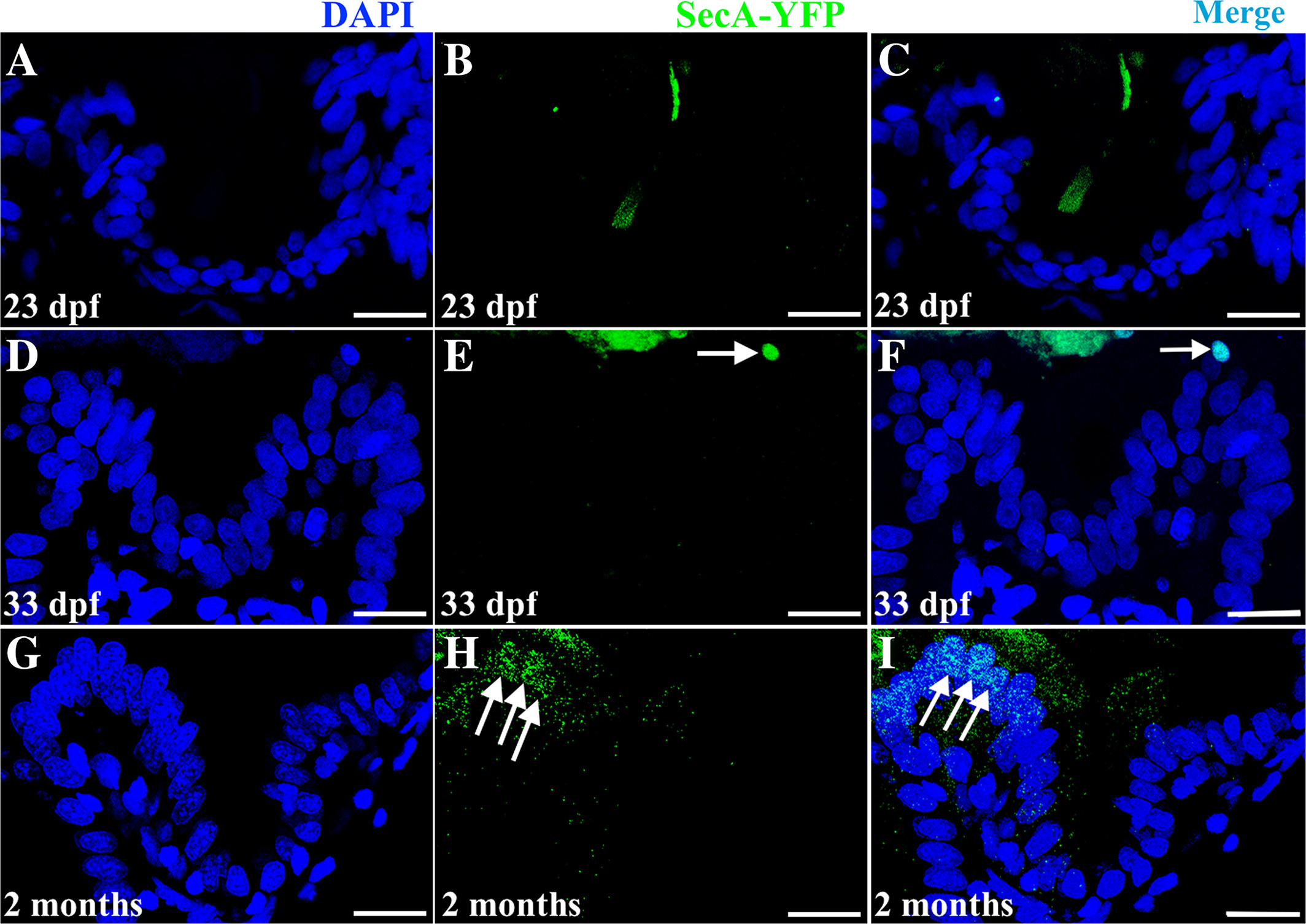
Fig. 10 A–I: Development of apoptotic cells. In cross sections of the Tg(βactin:SecAV‐YFP) pu17 line, no apoptotic cells are observed during the third postembryonic week (example 23 dpf A to C). A small numbers of apoptotic cells are first observed at the tip of the fold at 33 dpf (arrow in E and merge with DAPI nuclear stain in F). Apoptotic cells become more prominent at the fold tip by 2 months postembryogenesis (arrows in H and merge with DAPI nuclear stain in I).
Acknowledgments
This image is the copyrighted work of the attributed author or publisher, and
ZFIN has permission only to display this image to its users.
Additional permissions should be obtained from the applicable author or publisher of the image.
Full text @ Dev. Dyn.