
|
Fig. 10
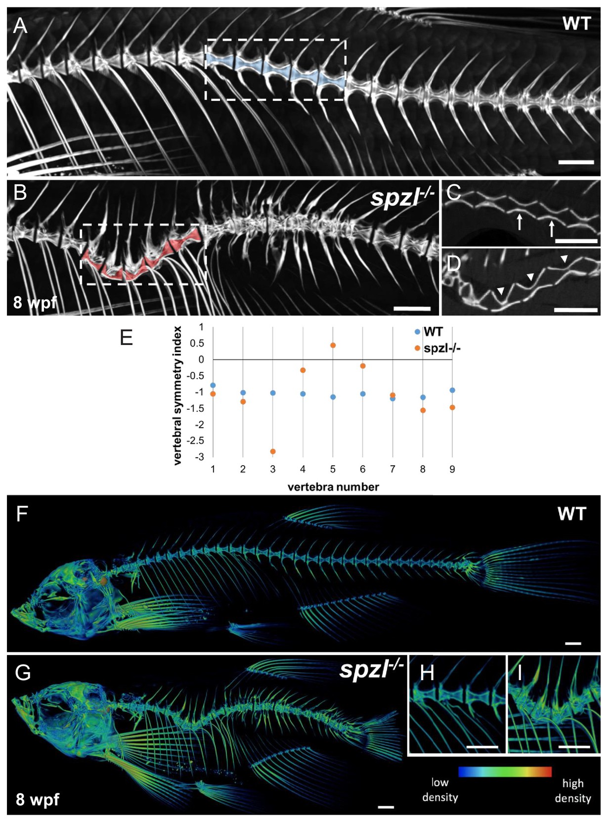
Vertebral malformations and increased mineralization in the spine of spzl-/- fish.
( A–B) μCT images of the spine of WT ( A) and spzl-/- ( B) fish at 8 wpf. Vertebral canals are highlighted in blue (WT) and red ( spzl-/-). ( C) Optical section of the dotted box in ( A) for WT. Arrows point to symmetrical vertebrae. ( D) Optical section of the dotted box in ( B). Arrowheads point to the vertebral asymmetry in spzl-/- associated with the spine kink. ( E) Vertebral symmetry index, calculated as in Figure 6E, of WT and spzl-/- obtained from the spine kinking area. Numbers correspond to vertebra number counted from the first rib bearing vertebra. ( F–G) μCT reconstruction of 6wpf WT and spzl-/- mutant zebrafish. The reconstructions were pseudo-colored to show relative bone density. ( H–I) Magnified insets of the spine for WT and spzl-/-. Scale bar = 1 mm.