Image
Figure Caption
Fig. 9-S1
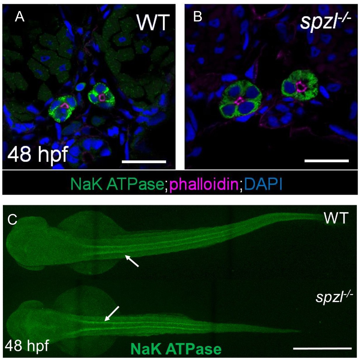
Pronephros development occurs normally in spzl mutants.
( A–B) Confocal images of cross sections from 48 hpf WT and spzl mutant embryos stained for NaK ATPase (green). Phalloidin (magenta) labels the apical membrane of the pronephros. Scale bar = 20 µm ( C) Whole mount immunofluorescence stain of NaK ATPase labeling thepronephros in WT (top) and spzl-/- (bottom). Arrows point to pronephros. Scale bar = 500 µm.
Acknowledgments
This image is the copyrighted work of the attributed author or publisher, and
ZFIN has permission only to display this image to its users.
Additional permissions should be obtained from the applicable author or publisher of the image.
Full text @ Elife

