
|
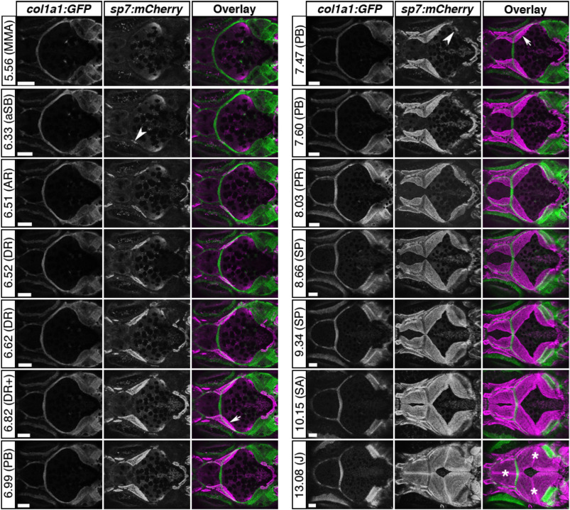
Fig. 3 Sequential live imaging of skull vault development. Confocal images are shown for a single Tg(col1a1:egfp; sp7:mcherry) zebrafish during the entire period of skull vault formation, with corresponding SL measurements in mm, and developmental staging in parentheses as in Fig. 2. Each set of panels represents maximum projections of z-stacks, separate GFP and mCherry channels and the overlay. Notable events are frontal bone initiation (arrowhead at 6.33 mm), planar bone growth towards apex of skull (arrows), parietal bone initiation (arrowhead at 7.47 mm), and suture formation (asterisks). Abbreviations as in Fig. 2, plus: metamorphic melanophore appearance (MMA); squamation onset posterior (SP); squamation through anterior (SA); juvenile (J). Scale bars = 100 μm.
Reprinted from Mechanisms of Development, 160, Kanther, M., Scalici, A., Rashid, A., Miao, K., Van Deventer, E., Fisher, S., Initiation and early growth of the skull vault in zebrafish, 103578, Copyright (2019) with permission from Elsevier. Full text @ Mech. Dev.