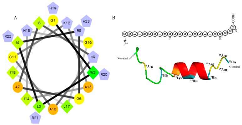
Predicted helical secondary and three-dimensional structures of Octominin. (A) Helical wheel of Octominin shows the AAs arrangement and the residue numbers which are counted from the amino (N) terminal of the peptide. The hydrophilic and hydrophobic residues are represented by circles and diamonds, respectively. The potentially charged (positively charged) residues are marked as pentagons in light blue. The most hydrophobic residue is green, and the amount of green is decreasing proportionally to the hydrophobicity, with zero hydrophobicity coded as yellow. (B) Three-dimensional structure and AA sequence of Octominin with positively charged residues.
Acknowledgments
This image is the copyrighted work of the attributed author or publisher, and
ZFIN has permission only to display this image to its users.
Additional permissions should be obtained from the applicable author or publisher of the image.
Full text @ Mar. Drugs
Your Input Welcome
Thank you for submitting comments. Your input has been emailed to ZFIN curators who may contact you if
additional information is required.
Oops. Something went wrong. Please try again later.