Image
Figure Caption

Figure 2

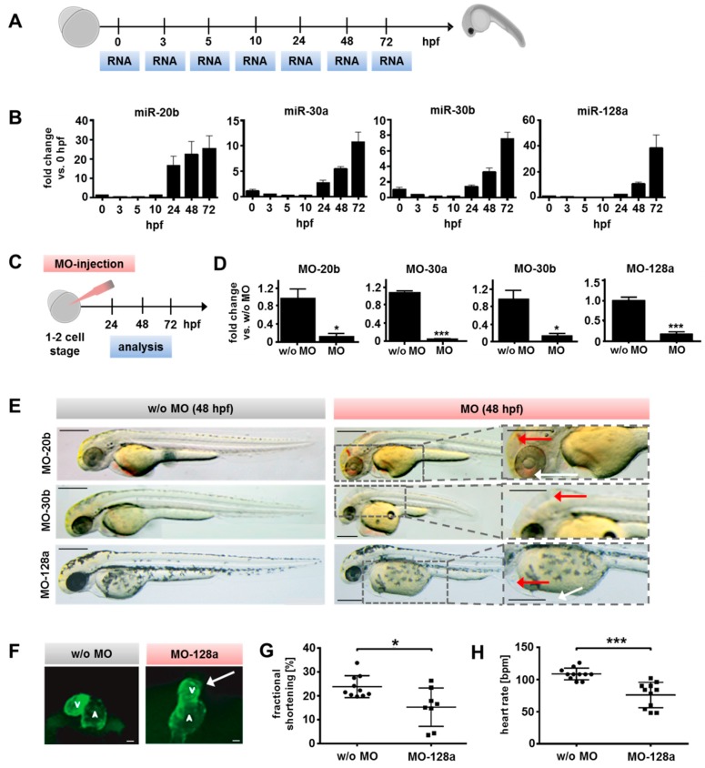
Evaluation of candidate miR function during zebrafish development. (A) MiR kinetics were evaluated during early zebrafish development. Total RNA was isolated on respective timepoints (hours post fertilization; hpf) for qRT-PCR. (B) MiR-20b, -30a, -30b, and -128a were upregulated in developing zebrafish larvae from 24 hpf increasing until 72 hpf. (C) Morpholino oligos (MOs) were injected in 1–2 cell-stage zebrafish embryos and miR knockdown, morphological changes, fractional shortening, and beating rate was analyzed (24, 48, 72 hpf). (D) MiR expression (evaluated by qRT-PCR) in zebrafish larvae was sufficiently reduced by MOs at 24 hpf by more than 85% for all candidate miRs in comparison to controls without MOs (t-test). (E) Morphological changes of MO-morphants at 48 hpf. MO-20b morphants showed cerebral hemorrhage (red arrow) and edema (edema in the eye region indicated by a white arrow). MO-30b morphants developed malformations and edemas (red arrow highlights a visible edema and enlarged hydrocephalus). MO-128a larvae exhibited a robust pericardial edema (red arrow) and blood congestion at the right outflow tract of the heart (white arrow). Scale bars: 250 µm. (F) Cardiac phenotype of MO-128a treated Tg(myl7:ras-GFP) zebrafish larvae at 48 hpf including smaller ventricles (white arrow) and abnormalities in heart looping. Scale bars: 5 µm. (G) MO-128a larvae (n = 8) showed a significantly reduced fractional shortening at 72 hpf compared to non-treated larvae (w/o MO, n = 10) (t-test) (H) MO-128a morphants (n = 11) appeared to have a significantly slower heart rate at 72 hpf compared to the controls (w/o MO, n = 11) (t-test). All data are represented as means ± SEM. * p ≤ 0.05, ** p ≤ 0.01 and *** p ≤ 0.001.

Figure Data
Acknowledgments
This image is the copyrighted work of the attributed author or publisher, and ZFIN has permission only to display this image to its users. Additional permissions should be obtained from the applicable author or publisher of the image. Full text @ Int. J. Mol. Sci.