
Figure 5
- ID
- ZDB-IMAGE-200306-30
- Publication
- Pang et al., 2020 - Mutant dlx3b disturbs normal tooth mineralization and bone formation in zebrafish
- All Figures
- Figures for Pang et al., 2020
|
Figure 5
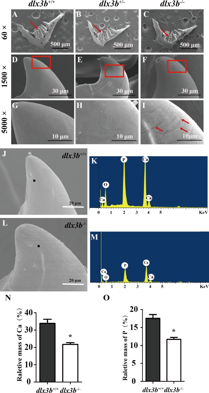
(A–I) SEM images showing the surface of the second pharyngeal tooth. The red arrows showed the teeth we studies below in a 60× image (A–C). The fourth ventral pharyngeal teeth (red arrows in A–C) was magnified as images D–F. The red rectangle areas in D–F were then magnified as G–I. The red arrows (I) indicate the pits on the surface of the second ventral pharyngeal tooth of the