IMAGE

Figure 5

ID
ZDB-IMAGE-200306-21
Source
Figures for Scheib et al., 2020
Image
Figure Caption

Figure 5

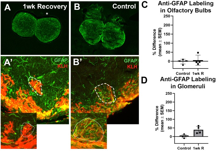
The recovery of the olfactory bulb does not involve evidence of a glial scar. (A) After the olfactory system was allowed 1 week to recover from 7 days of repetitive peripheral damage, there was no apparent difference in the overall amount of anti-GFAP (green) and anti-KLH (red) labeling in the affected bulb (asterisk) when compared to the internal control bulb (n = 4). There was also no noticeable difference in the overall amount of anti-GFAP labeling compared to untreated control fish (B). (A’) Higher magnification Z-stack images revealed that anti-GFAP labeling (green) within anti-KLH (red) labeled glomeruli (dotted line) in the affected bulb after 1 week of recovery also appeared to be similar to that of glomeruli in the internal control bulb (dotted outline in inset) and in untreated control bulbs (B’) on the right (dashed outline) and left (dotted outline in inset) sides. There was no significant difference in the percent difference of OD of anti-GFAP labeling in the olfactory bulb (C) or ventral medial glomeruli (D) of zebrafish that were allowed 1 week of recovery from 7 days of repetitive peripheral damage.

Acknowledgments
This image is the copyrighted work of the attributed author or publisher, and ZFIN has permission only to display this image to its users. Additional permissions should be obtained from the applicable author or publisher of the image. Full text @ Front. Neuroanat.