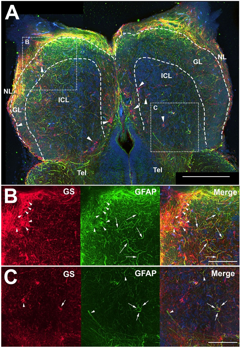
IMAGE

Figure 1

ID
ZDB-IMAGE-200306-13
Source
Figures for Scheib et al., 2020
Image
Figure Caption

Figure 1

Z-stack images of anti-glial fibrillary acidic protein (GFAP) and anti-glutamine synthetase (GS) immunoreactivity in the olfactory bulb of uninjured adult zebrafish. (A) There was a high degree of labeling for both antibodies in the nerve layer (NL) and glomerular layer (GL) of the olfactory bulb that varied in co-labeling. Anti-GS (red) labeling did not reveal clear structures with the exception of ring-like labeling seen in all layers of the bulb (arrowheads). There were numerous, distinct anti-GFAP (green) labeled processes in the GL and internal cell layer (ICL) that varied in size and length but had no obvious organization. Tel, Telencephalon. (B) Higher magnification of the rostral bulb region shown in (A) confirmed that ring-like structures (arrowheads) were labeled mostly with anti-GS and distinct processes (arrows) were labeled predominantly with anti-GFAP. (C) Higher magnification of the caudal bulb region revealed less labeling with both antibodies in this region, but anti-GS positive ring-like structures and anti-GFAP positive processes were apparent. Scale bar = 100 μm (A) or 20 μm (B,C).

Acknowledgments
This image is the copyrighted work of the attributed author or publisher, and ZFIN has permission only to display this image to its users. Additional permissions should be obtained from the applicable author or publisher of the image. Full text @ Front. Neuroanat.