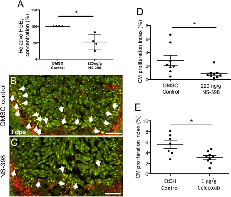
IMAGE

Figure 5

ID
ZDB-IMAGE-200302-32
Source
Figures for FitzSimons et al., 2020
Image
Figure Caption

Figure 5

Activation of the Cox2-PGE2 circuit stimulates cardiomyocyte proliferation. Zebrafish were subjected to ventricular amputation and treated with daily intraperitoneal (IP) injections of either vehicle control, NS-398 or Celecoxib. Hearts were collected for analysis at 3 dpa. (A) Treatment with NS-398 reduced PGE2 concentrations in ventricles by ~47%, relative to DMSO controls. (mean ± s.e.m. n = 4 biological replicate for each group; 6 pooled ventricles from weight-matched clutch mates per replicate. Student’s t-test. *P < 0.05). (B,C) Representative images of 3 dpa injured hearts treated daily with a DMSO control (B) or NS-398 (C). Hearts were stained with Mef2 (green), and Pcna (red). Mef2+Pcna+ cells mark proliferating CMs, highlighted by white arrows. Scale bars represent 50 μM. (D-E) CM proliferation indices were calculated as a percentage of Mef2(+)Pcna(+) cells relative to the total number of Mef2(+) cells in a defined area adjacent to the injury. (D) CM proliferation was reduced by more than 69% in animals treated with NS-398 when compared to controls. (E) Reduction of CM proliferation was greater than 54% in animals treated daily with Celecoxib, relative to controls. (mean ± s.e.m. n = 7–9 hearts from clutchmates; three sections were quantified per heart and results averaged. Student’s t-test. *P < 0.05).

Acknowledgments
This image is the copyrighted work of the attributed author or publisher, and ZFIN has permission only to display this image to its users. Additional permissions should be obtained from the applicable author or publisher of the image. Full text @ Sci. Rep.