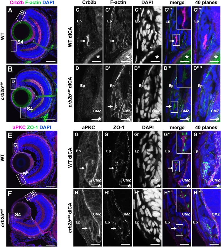
Crb2b is expressed in polarised cells of the ICA at 5 dpf. Immunostaining of transverse cryosections through the eye. (A,B,E,F) Overview of a WT (A,E) and a mutant (B,F) eye at 5 dpf. Higher magnifications of the boxed regions in the dorsal ICA are shown in C–D‴′ (WT) and G–H‴′ (crb2be40). Higher magnifications of the boxed regions in the ventral ICA are shown in Fig. S4. Crb2b staining (anti-Crb2be8e9) is detected in a cluster of cells in the WT ICA (C) but not in the mutant ICA (D). F-actin (C′,D′; visualised by phalloidin), aPKC (G,H) and ZO-1 (G′,H′) appear apically enriched (C′,D′). Arrows point to the cluster of polarised Crb2b expressing cells, and asterisks mark the lens. dICA, dorsal iridocorneal angle; Ep, epidermis; CMZ, ciliary marginal zone. Scale bars: (A,B,E,F) 50 µm; (C–D‴′, G–H‴′) 10 µm.
Acknowledgments
This image is the copyrighted work of the attributed author or publisher, and
ZFIN has permission only to display this image to its users.
Additional permissions should be obtained from the applicable author or publisher of the image.
Full text @ Biol. Open
Your Input Welcome
Thank you for submitting comments. Your input has been emailed to ZFIN curators who may contact you if
additional information is required.
Oops. Something went wrong. Please try again later.