IMAGE

Figure 5

ID
ZDB-IMAGE-200228-26
Source
Figures for Mei et al., 2019
Image
Figure Caption

Figure 5

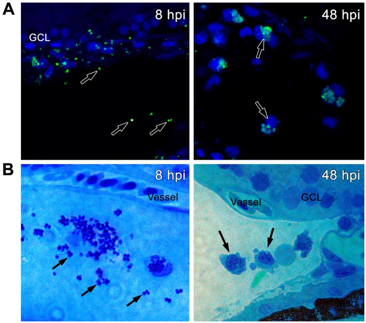
Zebrafish eyes (n = 10) were infected with GFP-positive S. aureus (250,000 CFU/eye). (A) Retinal cryosections were immunostained with anti-GFP antibody (green) and colabeled with TO-PRO 3 for nuclei (blue). S. aureus (as shown by black arrows) was located in the vitreous as well as in phagocytic cells. (B) Retinal sections were stained with Tetrazolium blue at indicated time points. The stained cells appeared to be macrophages with engulfed S. aureus in the vitreous cavity.

Acknowledgments
This image is the copyrighted work of the attributed author or publisher, and ZFIN has permission only to display this image to its users. Additional permissions should be obtained from the applicable author or publisher of the image. Full text @ Pathogens