
|
Fig. 2
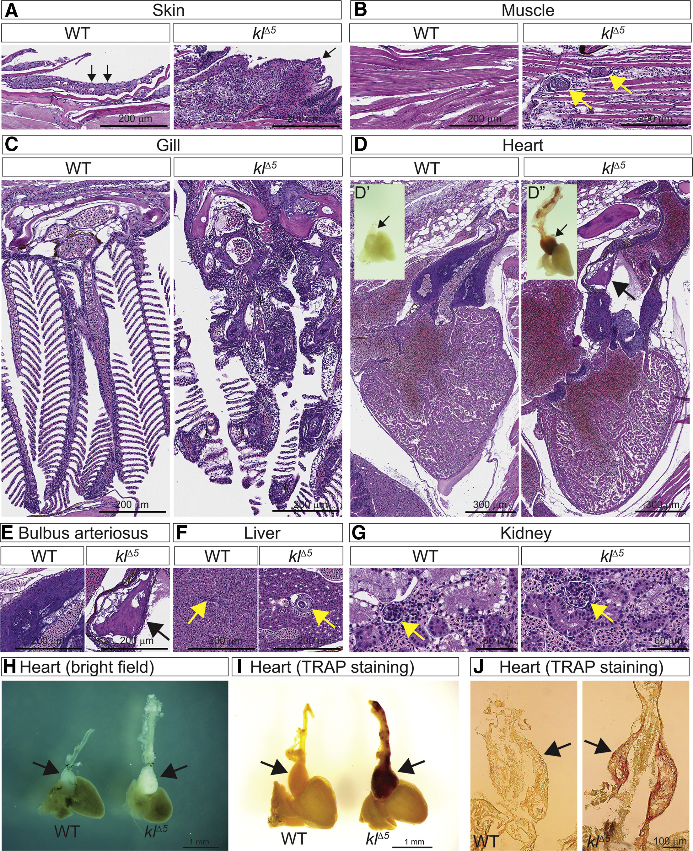
Vascular Calcification and Inflammation in αklothoMutants
H&E staining on paraffin sections from 5-month-old wild-type control (Tü) and αklotho (klΔ5) males. Shown are (A) skin (arrows indicate mucous cells in wild type); (B) muscle (arrows indicate vascular calcification); (C) gills; (D) heart (arrow indicates calcification in the BA); (D′ and D″) alizarin red-stained whole-mount hearts (arrows indicate the BA); (E) BA (arrow indicates calcification); (F) liver (arrows indicate bile-duct); and (G) kidney (arrows indicate glomeruli). (H) Bright-field images of 5-mpf wild-type (left) and klΔ5 (right) hearts. TRAP staining on (I) whole mount and (J) cryosection of 5-mpf hearts.