
Fig. 4
- ID
- ZDB-IMAGE-200210-64
- Publication
- Kossack et al., 2018 - Female Sex Development and Reproductive Duct Formation Depend on Wnt4a in Zebrafish
- All Figures
- Figures for Kossack et al., 2018
|
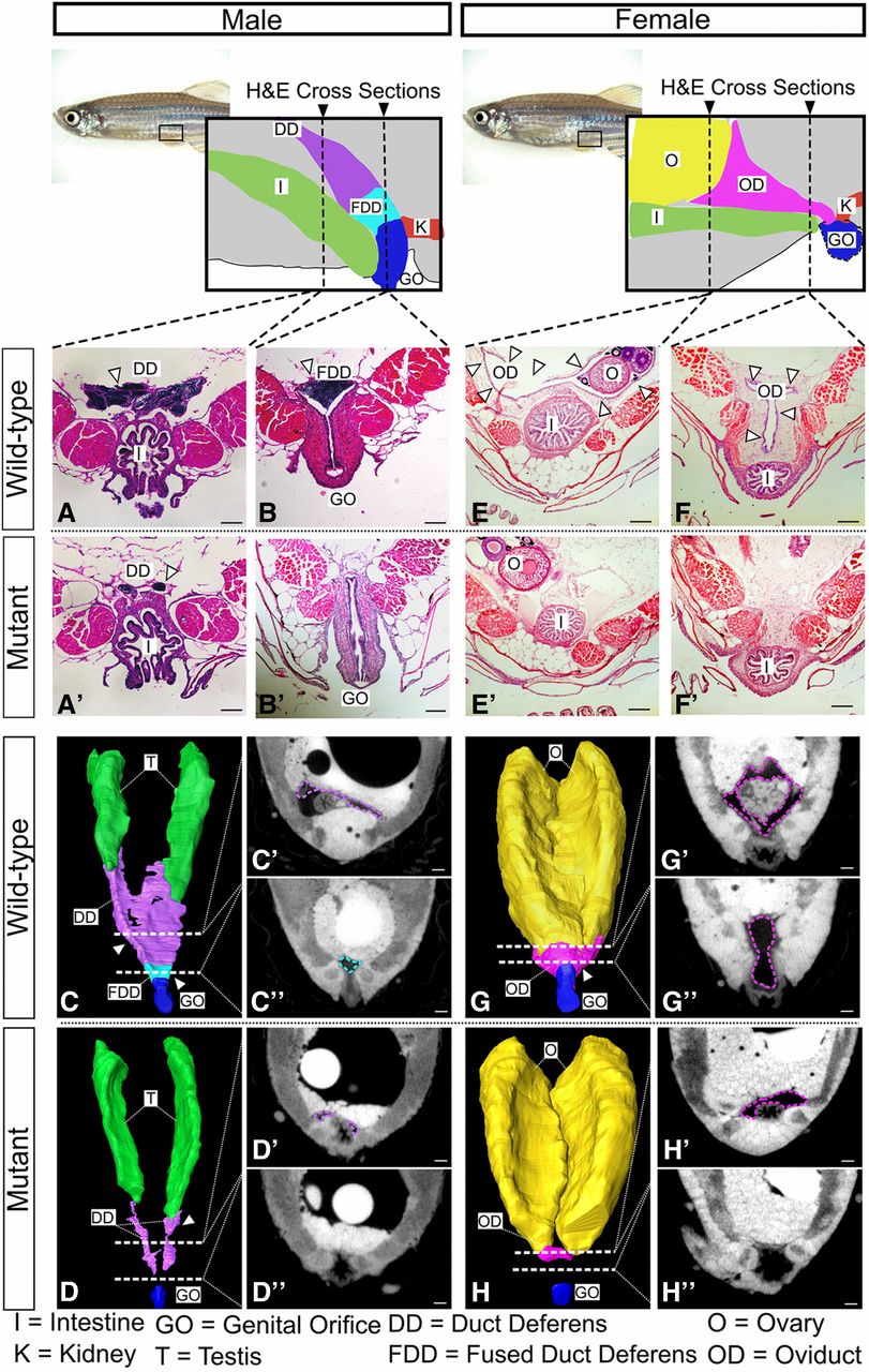
Fig. 4
Duct morphology in wnt4a mutant and wild-type males. From the posterior end of each testis, wild-type males developed duct deferens (A) that joined to form the fused duct deferens (B). Mutant males, however, failed to form a full duct deferens (A′) or a fused duct deferens (B′). Three-dimensional renderings built from individual traces of sections (C′, C″, D′, and D″) of (C) the wild-type ducts and (D) mutant ducts show that the mutant ducts never fully connected to the genital orifice. H&E-stained sections of wild-type females showed an oviduct that wrapped around the posterior of the ovaries (E, G, and G′) and extended ventroposteriorly (F, G, and G″) out to the genital orifice. Mutant females, however, failed to organize an oviduct around the ovary (E′, H, and H′) and did not form a connective duct to the genital orifice (F′, H, and H″). See movies in Files S1 and S2. Bar, 100 µm. I, intestine; K, kidney; T, testis, O, ovary; OD, oviduct; GO, genital orifice; DD, duct deferens; FDD fused duct deferens.