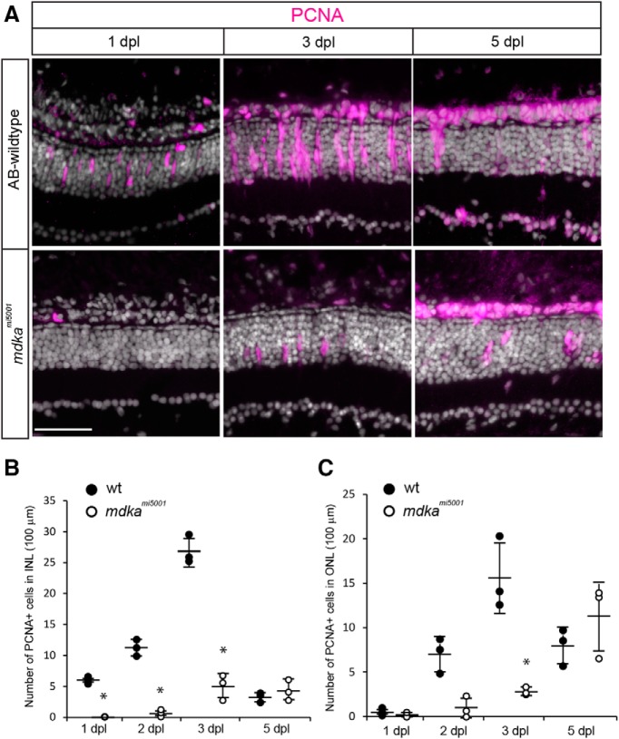
IMAGE

Figure 2.

ID
ZDB-IMAGE-200210-41
Source
Figures for Nagashima et al., 2019
Image
Figure Caption

Figure 2.

In the mdkami5001 mutant, Müller glia fail to proliferate in response to photoreceptor cell death. A, Immunocytochemistry for PCNA (magenta) in WT and mdkami5001 at 1 and 3 dpl. In WT, Müller glia become positive for PCNA at 1 dpl. At 3 dpl, PCNA+ progenitors form neurogenic clusters. Mutant retinas lack PCNA-labeled cells at 1 dpl. Very few, isolated cells are positive for PCNA in the mdkami5001 at 3 dpl. B, C, The number of PCNA+ cells in WT and mdkami5001 in the inner (B; 1 dpl: p = 0.0017; 2 dpl: p < 0.0001; 3 dpl: p < 0.0004, ANOVA with post hoc Tukey; F ratio = 116.1834) and outer (C; 3 dpl; p = 0.0001, ANOVA with post hoc Tukey; F ratio = 17.5589) nuclear layers. The mdkami5001 retinas have significantly less PCNA+ cells at 1, 2, and 3 dpl compared with WT retinas. A total of 3 sections were counted and averaged in each retina. A total of 3 retinas were analyzed. Scale bar, 30 μm. *p < 0.01.

Figure Data
Acknowledgments
This image is the copyrighted work of the attributed author or publisher, and ZFIN has permission only to display this image to its users. Additional permissions should be obtained from the applicable author or publisher of the image. Full text @ J. Neurosci.