
Fig. 2
- ID
- ZDB-IMAGE-200203-8
- Publication
- Zhu et al., 2019 - Zebrafish prmt5 arginine methyltransferase is essential for germ cell development
- All Figures
- Figures for Zhu et al., 2019
|
Fig. 2
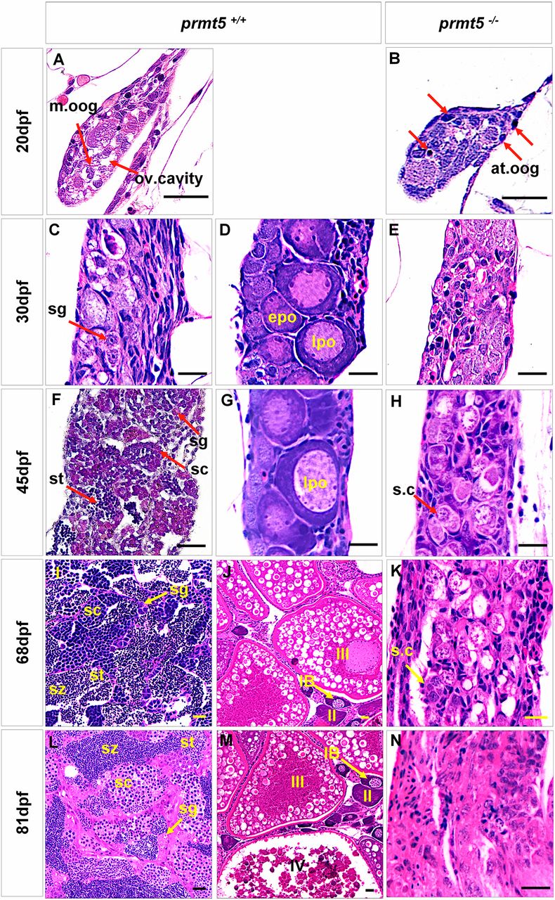
The gonads of prmt5−/− zebrafish fail to differentiate into normal testes or ovaries, as shown by histological comparisons of gonadogenesis. (A,B) At 20 dpf, prmt5−/− gonads contained a pyknotic mass and many atretic oogonia (at.oog, indicated by red arrows), while the prmt5+/+ gonads had a loosened structure with a prominent ovarian cavity (ov.cavity) and many mitotic oogonia (m.oog). (C-H) Between 30 and 45 dpf, the prmt5+/+ gonads began to differentiate into testes (C,F) or ovaries (D,G), as indicated by the presence of spermatogonia (sg), spermatocytes (sc) and spermatids (st), or by the increasing numbers of early stage IB perinucleolar oocytes(epo) and late stage IB perinucleolar oocytes (lpo). In contrast, the prmt5−/− gonads displayed a testis-like morphology containing only a few spermatogonia cysts (s.c). (I-N) Between 68 and 81 dpf, prmt5+/+ zebrafish possessed either mature testes (I,L), containing germ cells at different stages of spermatogenesis (spermatogonia, sg; spermatocytes, sc; spermatid, st; spermatozoa, sz), or mature ovaries (J,M), containing germ cells at different stages of oogenesis (IB, II, III or IV). The prmt5−/− gonads contained few spermatogonia cysts (s.c) (K) and instead contained several Sertoli cells (N). Scale bars: 20 μm; n=6 for each stage.