IMAGE

Fig. 3.

ID
ZDB-IMAGE-200130-23
Source
Figures for Lange et al., 2020
Image
Figure Caption

Fig. 3.

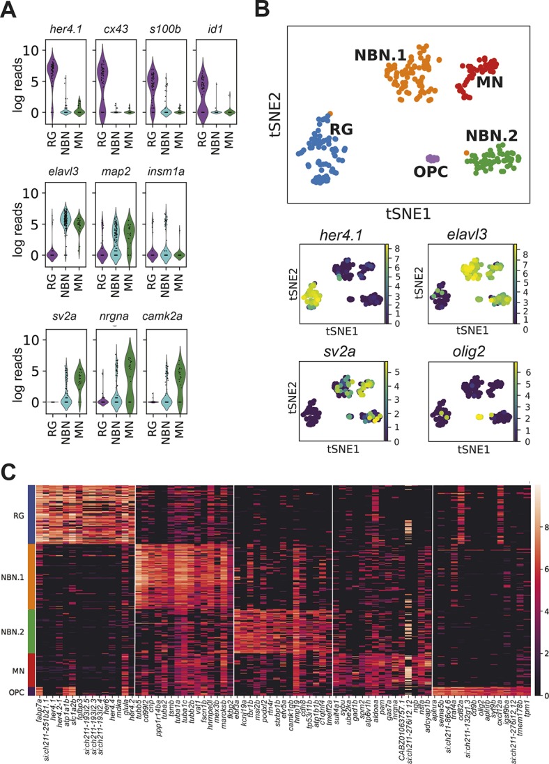
Single cell RNA-seq reveals the diversity of radial glia progeny in the adult forebrain. (A) Violin plots showing the expression of known radial glia markers (top), pan-neuronal markers (middle, elavl3 and map2), an early neurogenic fate marker (middle, insm1a) and markers of mature synaptically integrated neurons (bottom). (B) tSNE plot revealing five different clusters from a total of 264 RG, NBN and MN cells (top). The cell number per cluster is: RG, 76 cells; NBN.1, 80 cells; NBN.2, 54 cells; MN, 44 cells; and OPC, 10 cells. Smaller panels underneath show expression of characteristic marker genes that separate the different clusters. (C) Heat map with the top 15 cluster-specific genes for the five identified clusters.

Acknowledgments
This image is the copyrighted work of the attributed author or publisher, and ZFIN has permission only to display this image to its users. Additional permissions should be obtained from the applicable author or publisher of the image. Full text @ Development