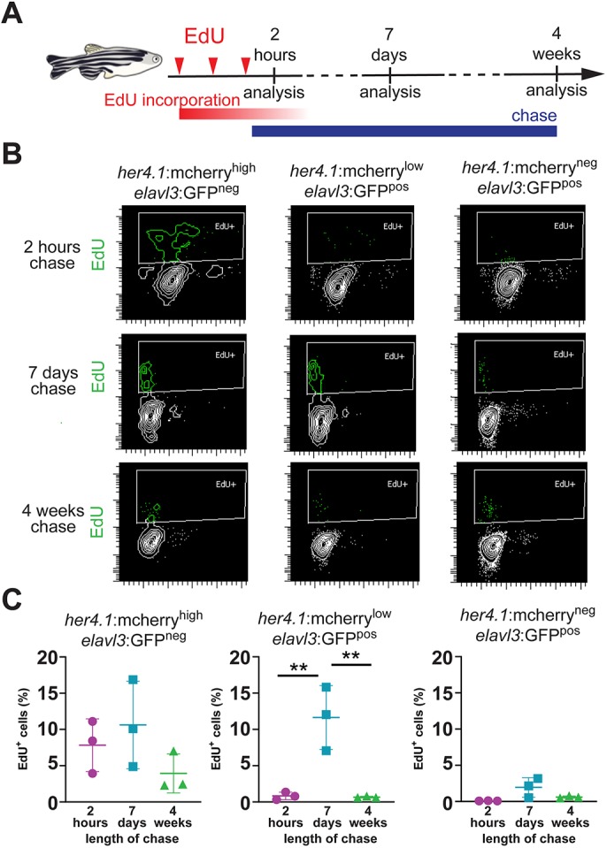
Newborn neurons retain the radial glia-derived reporter signal transiently. (A) Scheme of experimental design, indicating the timing of EdU injections (red arrowheads), EdU incorporation (red bar), EdU chase (blue bar) and analysis timepoints after the last EdU injection. (B) Flow cytometry plots showing EdU (green) incorporation in sorted her4.1:mCherryhigh/elavl3:GFPneg cells (left), her4.1:mCherrylow/elavl3:GFPpos cells (middle) and her4.1:mCherryneg/elavl3:GFPpos cells (right) after 2 h (top), 7 days (middle) or 4 weeks (bottom) of chase. There is robust EdU labeling in her4.1:mCherrylow/elavl3:GFPpos after 7 days, but not after 2 h or 4 weeks chase. (C) Quantification of EdU labeling her4.1:mCherryhigh/elavl3:GFPneg cells (left), her4.1:mCherrylow/elavl3:GFPpos cells (middle) and her4.1:mCherryneg/elavl3:GFPpos cells (right) after 2 h (magenta), 7 days (blue) or 4 weeks (green). Single data points are shown; n=3; data are mean±s.e.m. **P<0.01 by one-way ANOVA and Bonferroni's post hoc test.
Acknowledgments
This image is the copyrighted work of the attributed author or publisher, and
ZFIN has permission only to display this image to its users.
Additional permissions should be obtained from the applicable author or publisher of the image.
Full text @ Development
Your Input Welcome
Thank you for submitting comments. Your input has been emailed to ZFIN curators who may contact you if
additional information is required.
Oops. Something went wrong. Please try again later.