
Fig. 2
- ID
- ZDB-IMAGE-200124-53
- Publication
- Toms et al., 2019 - Missense variants in the conserved transmembrane M2 protein domain of KCNJ13 associated with retinovascular changes in humans and zebrafish
- All Figures
- Figures for Toms et al., 2019
|
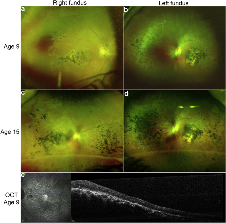
Fig. 2
Early clinical features of the
Ultra-wide field fundus imaging with Optos (Dunfermline, Scotland) of the right and left eye from patient A-2 with a missense mutation (p.Thr153Ile) in
Reprinted from Experimental Eye Research, 189, Toms, M., Dubis, A.M., Lim, W.S., Webster, A.R., Gorin, M.B., Moosajee, M., Missense variants in the conserved transmembrane M2 protein domain of KCNJ13 associated with retinovascular changes in humans and zebrafish, 107852, Copyright (2019) with permission from Elsevier. Full text @ Exp. Eye. Res.