
Fig. 1
- ID
- ZDB-IMAGE-200115-38
- Genes
- Publication
- Monestime et al., 2019 - CoRest1 regulates neurogenesis in a stage-dependent manner
- All Figures
- Figures for Monestime et al., 2019
|
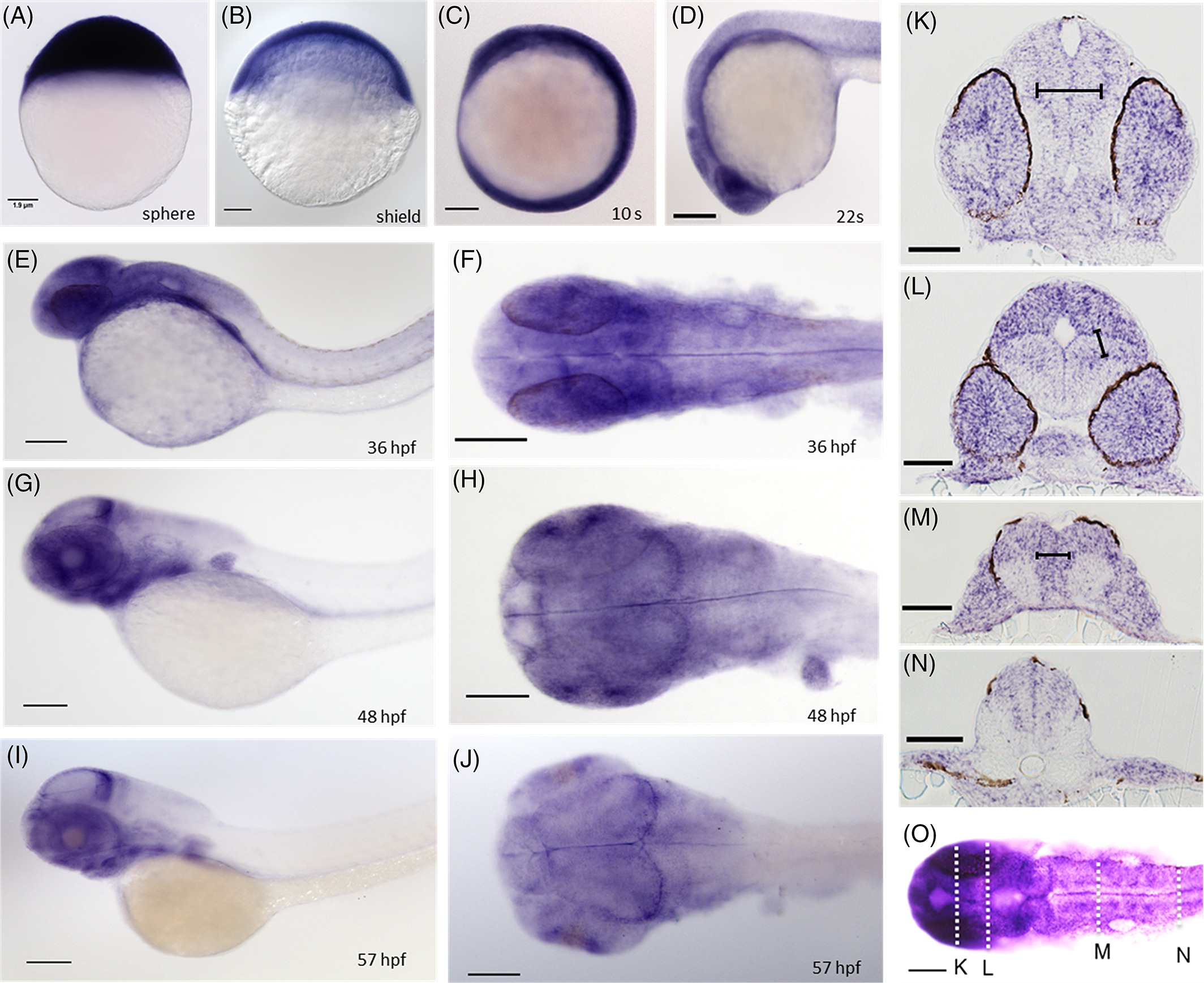
Fig. 1
Expression pattern of rcor1 in early zebrafish development. A‐C. At 4, 6, and 13 hpf rcor1 is ubiquitously expressed in the embryo. At 19 (D) and 36 hpf larvae have broad rcor1 expression throughout the head shown in both lateral and dorsal views at 36 hpf (E‐F). G‐J. Lateral and dorsal images of 48 and 57 hpf, respectively, show expression of rcor1 in the eye and posterior optic tectum. Transverse sections of 42 hpf larvae of the forebrain, midbrain, hindbrain, and anterior trunk (K‐N). Dorsal view of whole 42 hpf embryo used for transverse sectioning (O). Brackets outline the proliferative zone along the midline indicating regions of undifferentiated cell populations. Staining is seen in the eyes, OT, tg, and cg. hpf, hours postfertilization; OT, optic tectum; tg, tegmentum, cg, cranial ganglia (scale bar = 15 µm)