IMAGE

Figure 7

ID
ZDB-IMAGE-191230-969
Antibodies
Source
Figures for Liedtke et al., 2019
Image
Figure Caption

Figure 7

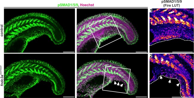
Investigation of TGF-beta/BMP signals in the median fin fold of fndc3awue1/wue1 mutants. Images show tail structures of representative control and fndc3awue1/wue1 mutant 22 hpf embryos stained for phosphorylated SMAD (pSMAD1/5/9) by immunofluorescence. Lack of pSMAD positive cells is observed in ventral median fin fold structures in fndc3awue1/wue1 mutants. All pictures display maximum intensity projections. White arrows indicate loss of pSMAD signals. Fire LUT shows pseudo-colored pSMAD1/5/9 signals. Dashed white lines indicate median fin fold borders. Scale bars: 50 µm.

Figure Data
Acknowledgments
This image is the copyrighted work of the attributed author or publisher, and ZFIN has permission only to display this image to its users. Additional permissions should be obtained from the applicable author or publisher of the image. Full text @ Sci. Rep.