IMAGE

Fig. 3

ID
ZDB-IMAGE-191230-947
Genes
Antibodies
Source
Figures for Hughes et al., 2019
Image
Figure Caption

Fig. 3

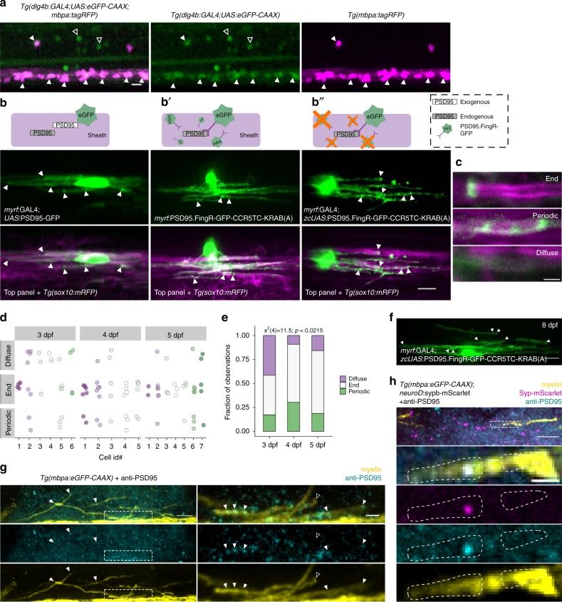
PSD95 is expressed by myelinating oligodendrocytes and is variably localized within myelin sheaths. a CRISPR/Cas9-mediated GAL4 enhancer trap of dlg4b reports spinal cord cells that express dlg4b via UAS:eGFP-CAAX expression. Larvae additionally carrying the mbpa:tagRFP transgene reveal mbp+ myelinating oligodendrocytes. Closed arrowheads indicate mbp+, dlg4b+ oligodendrocytes and open arrowheads indicate mbp-, dlg4b+ cells. Scale bar, 10 µm. b, b′, b″) PSD95 localization within oligodendrocytes detected by expression of exogenous PSD95-GFP fusion protein (b), expression of PSD95.FingR to detect endogenous PSD95 localization (b′), and expression of PSD95.FingR to detect endogenous PSD95 localization with a transcriptional repression system to limit unbound PSD95.FingR-GFP (b″). Arrowheads indicate puncta, scale bar 10 µm. c Expression of the transcriptionally-regulated system (b″) with Tg(sox10:mRFP) to label myelin shows that not all sheaths display localized PSD95 puncta (“diffuse”), and among those that do, labeling is either restricted to the sheath end (“end”), or periodic along the length of the sheath (“periodic”). Scale bar, 2 µm. d Classification plot of sheath labeling patterns (y-axis) for individual cells (x-axis) at 3, 4, and 5 dpf. n = 6 cells/29 sheaths (3 dpf); 5 cells/33 sheaths (4 dpf); 7 cells/32 sheaths (5 dpf). For each age group, dots of the same color denote sheaths belonging to the same cell. Note that most cells formed sheaths with different labeling patterns. Over developmental time, the distribution of labeling patterns changes as assessed by Chi square test (e), notably with a reduction of diffuse labeled sheaths, but all three categories persist at 5 dpf. (f) Later in development (8 dpf), PSD95.FingR-GFP is less punctate overall but remains enriched at the ends of sheaths (arrowheads). g Sagittal section of a 5 dpf Tg(mbpa:eGFP-CAAX) larva spinal cord fixed and immunolabeled with anti-PSD95. Right panels show higher magnification of the boxed region. Arrowheads indicate PSD95 signal colocalized with sheaths; open arrowhead marks a sheath without PSD95 at the terminal end. h Similar to g but with transient expression of neuroD:sypb-mScarlet to sparsely label synaptic vesicle puncta. Inset shows colocalized PSD95 and Syp-mScarlet within a sheath. Scale bars are 10 µm and 2 µm (insets)

Figure Data
Acknowledgments
This image is the copyrighted work of the attributed author or publisher, and ZFIN has permission only to display this image to its users. Additional permissions should be obtained from the applicable author or publisher of the image. Full text @ Nat. Commun.