
Fig. 2
- ID
- ZDB-IMAGE-191230-825
- Publication
- Thestrup et al., 2019 - A morphogenetic EphB/EphrinB code controls hepatopancreatic duct formation
- All Figures
- Figures for Thestrup et al., 2019
|
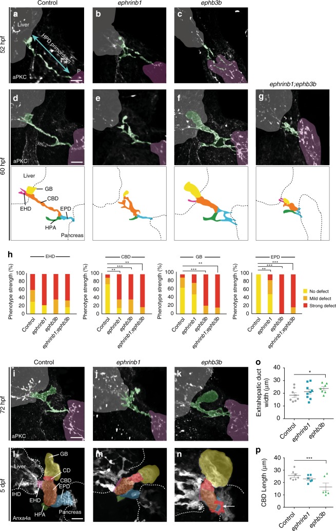
Fig. 2
EphrinB1 and EphB3b control HPD remodeling in a spatiotemporal fashion. Compared to sibling controls at 52 hpf (