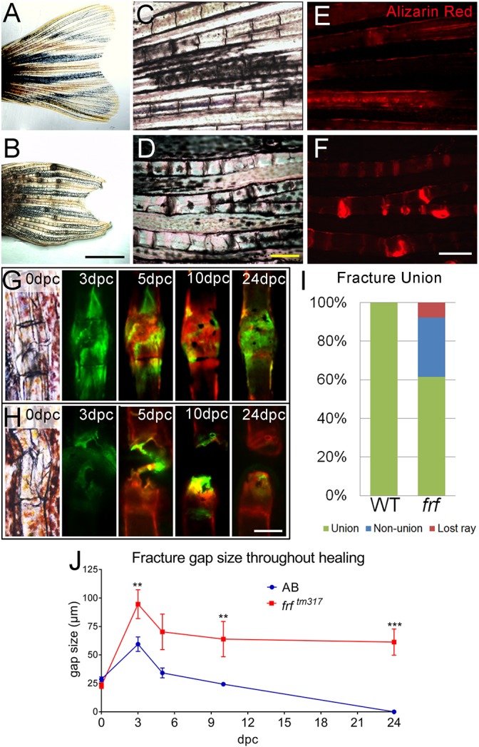
Non-union fractures in a zebrafish OI model. (A-D) Brightfield images at low (A,B) and high (C,D) magnification of adult fins at 3 months post fertilisation, showing thickening and dark foci in frf mutants (B,D) compared with WT (A,C). (E,F) Magnified images of WT (E) and frf (F) foci stained with Alizarin Red demonstrates bone calluses at these sites, indicating fractures. (G,H) Brightfield and fluorescent images of induced fractures in WT (G) and frf (H) at the indicated dpc. Bone was alternately stained with calcein (green) or Alizarin Red (red) to visualise callus growth. (I) A proportion of induced fractures in frf were non-union; n=12 per genotype. (J) The gap size following fracture was not reduced compared with WT over time; n=12 per point. **P<0.01, ***P<0.001; ANOVA with Sidak post test. Scale bars: in B, 2 mm for A,B; in D and F, 200 µm for C-F; in H, 100 µm for G,H.
This image is the copyrighted work of the attributed author or publisher, and
ZFIN has permission only to display this image to its users.
Additional permissions should be obtained from the applicable author or publisher of the image.
Full text @ Dis. Model. Mech.
Your Input Welcome
Thank you for submitting comments. Your input has been emailed to ZFIN curators who may contact you if
additional information is required.
Oops. Something went wrong. Please try again later.