
|
Figure 6
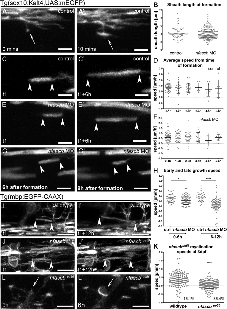
Neurofascin B Regulates Myelin Sheath Elongation and Stability
(A and A′) Single time-point confocal images of a myelinating process in a Tg(sox10KalTA4, UAS mEGFP) control that makes contact with a target axon (A) and transitions into a recognizable myelin sheath (A′). Scale bar, 5 μm.
(B) Average length of myelin sheaths immediately upon formation in control and
(C and C′) Confocal images of a myelin sheath elongating over time during the period of sheath formation in a control animal. Arrowheads in all panels denote ends of myelin sheaths. Scale bar, 5 μm.
(D) Average speed of individual sheath elongation in controls over a 12-h period, starting from the hour of their formation during the critical period. Each point represents a single sheath, imaged across 13 animals. No significant difference in speed of growth based on time of sheath formation, p = 0.8933, Kruskal-Wallis test.
(E and E′) Single time-point confocal images of a myelin sheath elongating over time during the critical period of sheath formation in an
(F) Average speed of sheath elongation in
(G and G′) Single time-point confocal images of a myelin sheath shrinking over time after the critical period in a
(H) Speed of elongation of individual myelin sheaths in control and
(I and I′) Confocal images of a myelin sheath elongating in a wildtype animal. Scale bar, 10 μm.
(J and J′) Confocal images of a myelin sheath elongating in an
(K) Speed of myelin sheath elongation in Tg(mbp:EGFP-CAAX) wildtype and
(L) Cell body myelination in
Reprinted from Developmental Cell, 51(6), Klingseisen, A., Ristoiu, A.M., Kegel, L., Sherman, D.L., Rubio-Brotons, M., Almeida, R.G., Koudelka, S., Benito-Kwiecinski, S.K., Poole, R.J., Brophy, P.J., Lyons, D.A., Oligodendrocyte Neurofascin Independently Regulates Both Myelin Targeting and Sheath Growth in the CNS, 730-744.e6, Copyright (2019) with permission from Elsevier. Full text @ Dev. Cell