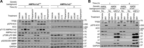
TFEB translocates to the nucleus upon AMPK stimulation independently of mTOR in mouse primary hepatocytes. A) AMPKα1α2+/+ or AMPKα1α2−/− mouse primary hepatocytes were isolated and cells were treated with 10 µM 991, 300 µM AICAR, 30 µM C13, or rapamycin 0.05 µM for 1 or 4 h. Western blot analysis of the indicated proteins was performed on cell lysates. B) Primary hepatocytes were isolated from AMPKα1α2−/− mice and were untreated (No) or coinfected with 3 adenoviruses (1:3 multiplicity of infection) in order to rescue the expression of the 3 subunits of AMPK (Flag-α2, β1, and γ1). Sixteen hours postinfection, the primary hepatocytes were treated for 1 h with vehicle (DMSO) or 10 µM 991. Cell lysates (20 µg) were used for Western blot analysis with the indicated antibodies. Images are representative of n = 3.
Acknowledgments
This image is the copyrighted work of the attributed author or publisher, and
ZFIN has permission only to display this image to its users.
Additional permissions should be obtained from the applicable author or publisher of the image.
Full text @ FASEB J.
Your Input Welcome
Thank you for submitting comments. Your input has been emailed to ZFIN curators who may contact you if
additional information is required.
Oops. Something went wrong. Please try again later.