
Figure 9
- ID
- ZDB-IMAGE-191230-1437
- Publication
- Duchemin et al., 2019 - Mechanically activated Piezo channels modulate outflow tract valve development through the Yap1 and Klf2-Notch signaling axis
- All Figures
- Figures for Duchemin et al., 2019
|
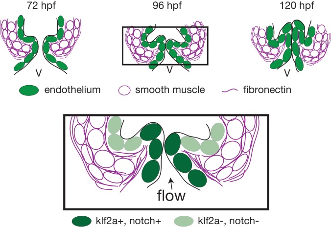
Figure 9 Working model summarizing OFT valve morphogenesis in response to mechanical forces. Two cell layers forming the OFT respond to piezo1 activity: the endothelium (green) and smooth muscle cells (magenta). Endothelial klf2a expression is repressed by Piezo1. In the smooth muscle cells, the expression of elastin (Elnb), fibronectin (Fn1) and Yap1 is modulated by Piezo1. Fibronectin is localized in the smooth muscle cell layer and within the forming valve (magenta lines). Future work will help to decipher how the two cell layers interact with each other to modulate OFT valve formation. V, ventricle.