
|
Figure 1
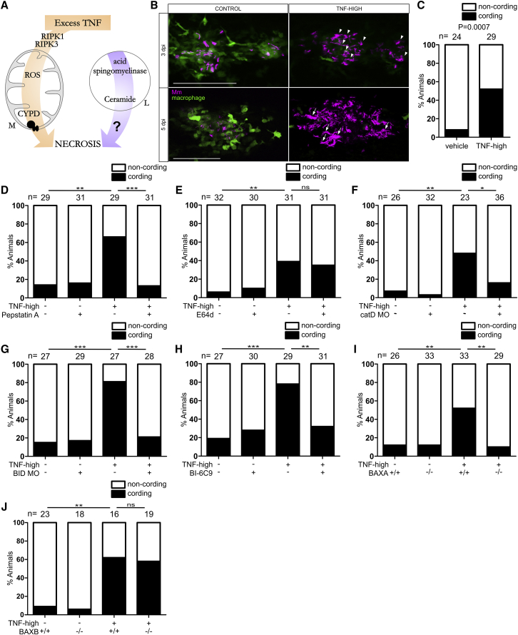
Ceramide Causes Necrosis through Cathepsin D, BID, and BAX
(A) Cartoon of TNF-mediated necrosis pathway components. CYPD, cyclophilin D; M, mitochondrion; L, lysosome.
(B) Confocal images of granulomas in 3 or 5 dpi TNF-high or control larvae with yellow fluorescent macrophages infected with red fluorescent Mm. Arrowheads, extracellular bacteria; arrows, extracellular, cording bacteria. Scale bar, 100 μm.
(C) Cording in 5 dpi TNF-high and control larvae.
(D) Cording in 5 dpi TNF-high or control larvae treated with pepstatin A.
(E) Cording in 5 dpi TNF-high or control larvae treated with E64d.
(F) Cording in 5 dpi TNF-high and control larvae that are wild-type (WT) or cathepsin D morphant.
(G) Cording in 5 dpi TNF-high and control larvae that are WT or BID morphant.
(H) Cording in 5dpi TNF-high and control larvae treated with BI-6C9.
(I) Cording in 5 dpi TNF-high or control larvae that are WT or BAXA mutant.
(J) Cording in 5 dpi TNF-high and control larvae that are WT or BAXB mutant.
(C–J) ∗p < 0.05; ∗∗p < 0.01; ∗∗∗p < 0.001 (Fisher’s exact test). Each panel representative of 3–6 independent experiments.
See also
Reprinted from Cell, 178(6), Roca, F.J., Whitworth, L.J., Redmond, S., Jones, A.A., Ramakrishnan, L., TNF Induces Pathogenic Programmed Macrophage Necrosis in Tuberculosis through a Mitochondrial-Lysosomal-Endoplasmic Reticulum Circuit, 1344-1361.e11, Copyright (2019) with permission from Elsevier. Full text @ Cell