IMAGE

Figure 4

ID
ZDB-IMAGE-191230-1239
Source
Figures for Jin et al., 2019
Image
Figure Caption

Figure 4

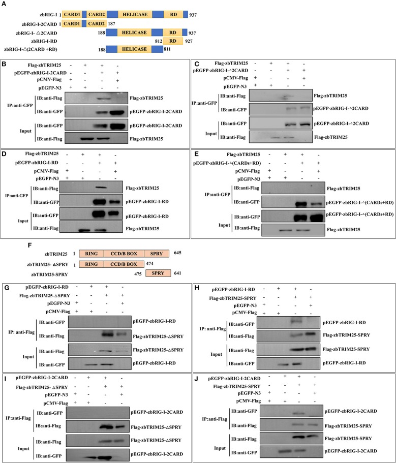
Physical interaction of zbTRIM25 with zbRIG-I. (A) Schematic representation of full-length zbRIG-I and zbRIG-I deletion mutants. (B–E) Interactions of GFP-tagged zbRIG-I-2CARD (B), zbRIG-I-Δ2CARD (C), zbRIG-I-RD (D), or zbRIG-I-Δ(CARDs+RD) (E) with Flag-tagged zbTRIM25 were examined using immunoprecipitation assays. HEK 293T cells were transfected with plasmids as indicated for 24 h. After treated with poly I:C for 24 h, cells were lysed, and the cell lysates were either analyzed directly by using anti-GFP and anti-Flag antibodies via Western blotting (Input) or subjected to immunoprecipitation using anti-GFP antibodies. The precipitates (IP) were analyzed by Western blotting with anti-GFP and anti-Flag antibodies, respectively. (F) Schematic illustration of zbTRIM25 truncations. Interactions of GFP-zbRIG-I-RD with Flag-zbTRIM25-ΔSPRY (G) or Flag-zbTRIM25-SPRY (H), GFP-zbRIG-I-2CARD with Flag-zbTRIM25-ΔSPRY (I), or Flag-zbTRIM25-SPRY (J) were examined using immunoprecipitation assays. HEK 293T cells were transfected with plasmids as indicated for 24 h. Cells were lysed after treated with poly I:C for 24 h. Immunoprecipitation and immunoblotting were performed with indicated antibodies.

Acknowledgments
This image is the copyrighted work of the attributed author or publisher, and ZFIN has permission only to display this image to its users. Additional permissions should be obtained from the applicable author or publisher of the image. Full text @ Front Immunol