IMAGE

Figure 6.

ID
ZDB-IMAGE-191230-1218
Source
Figures for Ge et al., 2019
Image
Figure Caption

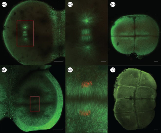
Figure 6.

Immunohistochemistry of zebrafish embryos during early cleavage in control condition. (a) Metaphase of the second cleavage, (b) magnification of the spindle in a, (c) embryo of 4-cell stage after the second cleavage, (d) telophase of the third cleavage, (e) magnification of the departing spindle in (d) and (f) embryo of 8-cell stage after the third cleavage and before the telophase of the fourth cleavage. Green indicates microtubules and red indicates chromosomes. Scale bar in a, c, d and f is 50 µm. Scale bar in b and e is 10 µm.

Acknowledgments
This image is the copyrighted work of the attributed author or publisher, and ZFIN has permission only to display this image to its users. Additional permissions should be obtained from the applicable author or publisher of the image. Full text @ Open Biol.