Image
Figure Caption

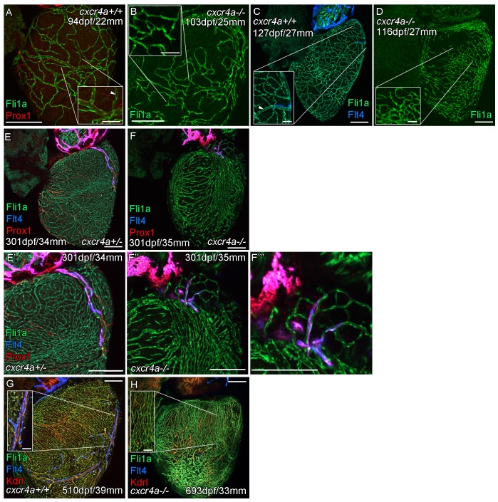
Figure 3—figure supplement 1. Cardiac lymphatic vessels extension is correlated with extent of the coronary arterial tree.

Whole-mount confocal imaging of adult transgenic zebrafish hearts expressing the pan-endothelial fli1a:GFP (green: A-H), Arterially and endocardially enriched endothelial marker kdrl:mTurquoise (red: G and H), lymphatic expressed flt4:mCitrine (blue: C, E-H); prox1:Gal4-UAS:RFP (red: A, E, F). (A and C) Formation of coronary vessels prior to and during the extension of the lymphatic vessel onto the ventricle in wildtype transgenic zebrafish (arrow head: lumenized vessel, A; coronary artery, B). (B) Much of the coronary vasculature is not formed in adult cxcr4a mutant zebrafish prior to the lymphatic extension and there are no identifiable lumenized vessels. (D) The formation of some coronary vessels in older adult mutants is mispatterned and abnormal during the time of lymphatic extension. (E and F) Images of hearts showing mispatterned coronary vasculature and failure of lymphatic extension in 301dpf cxcr4a mutants (E’, F’’ and F’’’ higher magnification images of the respective lymphatic vessels). (G and H) Lack of cardiac lymphatic vessels on the ventricle persists in the majority of older adult cxcr4a mutants (693dpf) that have considerably more coronary vessels than at earlier stages. (H) 693dpf cxcr4a mutant zebrafish lack identifiable coronary arteries that normally express high levels of kdrl:mTurquoise and along which flt4:mCitrine positive lymphatic vessels align (G, inset). Scale bars 200 μm and 50 μm (insets A-D, G and H).

Acknowledgments
This image is the copyrighted work of the attributed author or publisher, and ZFIN has permission only to display this image to its users. Additional permissions should be obtained from the applicable author or publisher of the image. Full text @ Elife