
Fig. S6
- ID
- ZDB-IMAGE-191018-5
- Publication
- Espenschied et al., 2019 - Epithelial delamination is protective during pharmaceutical-induced enteropathy
- All Figures
- Figures for Espenschied et al., 2019
|
Fig. S6
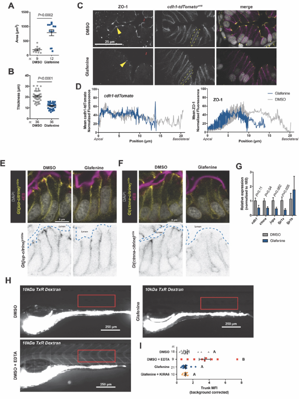
Epithelial junctional organization is overtly normal and barrier function is preserved in Glafenine-treated zebrafish larvae.
(A,B) Morphometric analysis of intestinal lumen area and epithelial thickness from transverse sections of 6 dpf larvae. Significance determined by unpaired two-sided Student’s t-test.
(C) Confocal micrographs of transverse vibratome sections from DMSO and Glafenine-treated cdh1-
tdTomatoxt18 (magenta) larvae stained with anti ZO-1 (yellow) and DAPI (gray). Yellow arrowheads point to apical ZO-1 staining and yellow dashed ovals denote representative basolateral patterning of Cdh1- tdTomato.
(D) Representative traces of Cdh1-tdTomato and ZO-1 localization patterns from vibratome sections of larvae treated as indicated.
(E,F) Representative confocal images of vibratome sections from 6 dpf DMSO and Glafenine treated Gt(jup-citrine)ct520a and Gt(ctnna-citrine)ct3a larvae (upper panels show merged images including DAPI and the brush border marker 4E8, lower panels show inverted grayscale images of indicated gene trap . lines).
(G) qRT-PCR analysis of indicated epithelial junctional components from FACS-isolated fabp2:DsRed+ enterocytes. Significance determined by unpaired two-sided Student’s t-test.
(H) Representative images of larvae 30 minutes post gavage with 10 kDa Texas Red dextran (DMSO larvae co-gavaged with EDTA were used as a positive control group for barrier permeability).
(I) Quantification of fluorescence in the trunk from larvae treated as indicated (example ROI shown in red boxes in H). Significance determined by one-way ANOVA with Tukey’s multiple comparison’s test; letters indicate groups determined to be statistically different.