
Fig. S3
- ID
- ZDB-IMAGE-191018-4
- Publication
- Espenschied et al., 2019 - Epithelial delamination is protective during pharmaceutical-induced enteropathy
- All Figures
- Figures for Espenschied et al., 2019
|
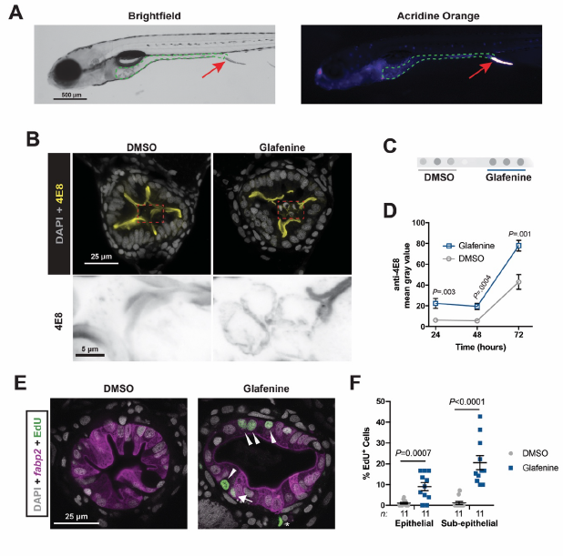
Fig. S3
Expelled mass of apoptotic cells contains enterocytes, and Glafenine treated larvae exhibit increased intestinal proliferation.
(A) Representative brightfield and AO fluorescence images of a 6 dpf Glafenine-treated zebrafish larva excreting AO+ IEC mass.
(B) Representative confocal micrographs of transverse sections immunostained with the IEC brush border antibody 4E8 (merge with DAPI above, inverted 4E8 below).
(C,D) Representative media dot blot of one sample pair probed with anti-4E8 and quantification (n = 4 samples / condition / time point, blotted in triplicate). (E,F) Analysis of epithelial proliferation in Tg(fabp2:DsRed) DMSO- and Glafenine-treated larvae (arrow heads mark EdU+ enterocytes, arrow denotes EdU+ sub-epithelial cell, * EdU+ extra-intestinal cell; significance determined by unpaired two-sided Student’s t-test).