
Fig. 5
- ID
- ZDB-IMAGE-191014-5
- Publication
- Mathewson et al., 2019 - Microtubules are required for the maintenance of planar cell polarity in monociliated floorplate cells
- All Figures
- Figures for Mathewson et al., 2019
|
Fig. 5
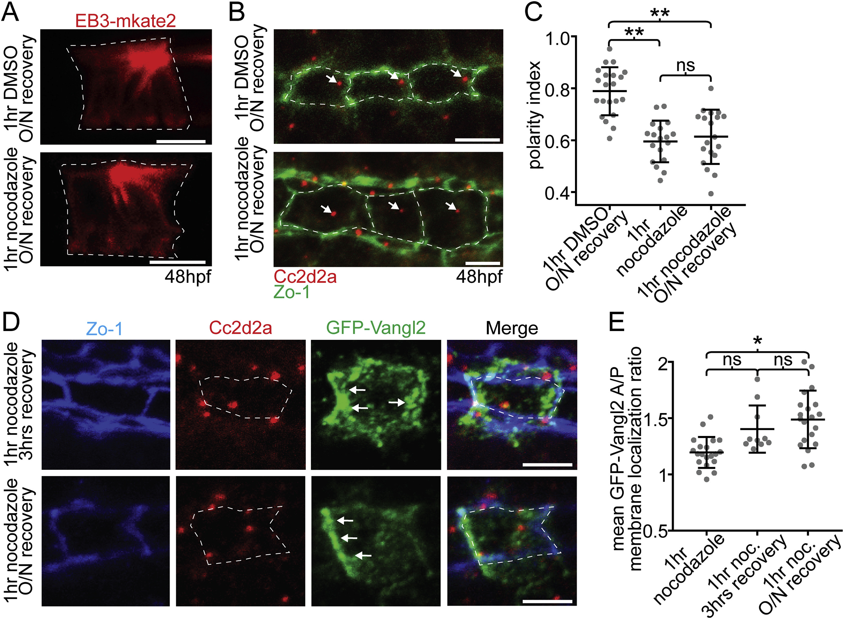
Maintenance of PCP protein polarity does not depend on a polarized BB. (A) EB3-mkate localization to centrosomal MTs recovers after recovery from nocodazole treatment (compare to Fig. 4A). (B) Ventral images of fixed 48hpf WT floorplates coimmunostained for Cc2d2a (red) and ZO-1 (green). BBs (arrows) remain delocalized after recovery from nocodazole treatment. (C) Quantitation of per embryo average BB polarity index in WT embryos that were treated with either DMSO (control) or cold 5 ng/μl nocodazole for 1hr and then either fixed immediately or allowed to recover overnight. N = 615 cells, 58 embryos; **p < 0.0001; significance was determined with a Kruskal-Wallis test with Dunn's multiple comparison. (D) Ventral images of fixed GFP-Vangl2 expressing floorplate cells in Tg(shh:gal4); Tg(uas:GFP-Vangl2) embryos that were treated with nocodazole for 1hr and then recovered for either 3 hrs or overnight. Arrows indicate recovery of asymmetric GFP-Vangl2 localization. Compare to Fig. 4D. (E) Quantitation of GFP-Vangl2 anterior vs. posterior membrane localization ratios after 1hr nocodazole treatment followed by different recovery periods. N = 236 cells, 29 embryos; *p = 0.0004; significance was determined with a Kruskal-Wallis test with Dunn's multiple comparison. White dotted lines mark approximate cell boundaries, based on ZO-1 staining of tight junctions. Scale bars: 5 μm.
Reprinted from Developmental Biology, 452(1), Mathewson, A.W., Berman, D., Moens, C.B., Microtubules are required for the maintenance of planar cell polarity in monociliated floorplate cells, 21-33, Copyright (2019) with permission from Elsevier. Full text @ Dev. Biol.