
Fig. 3
- ID
- ZDB-IMAGE-191007-2
- Publication
- Grassini et al., 2019 - Myosin Vb is required for correct trafficking of N-cadherin and cardiac chamber ballooning
- All Figures
- Figures for Grassini et al., 2019
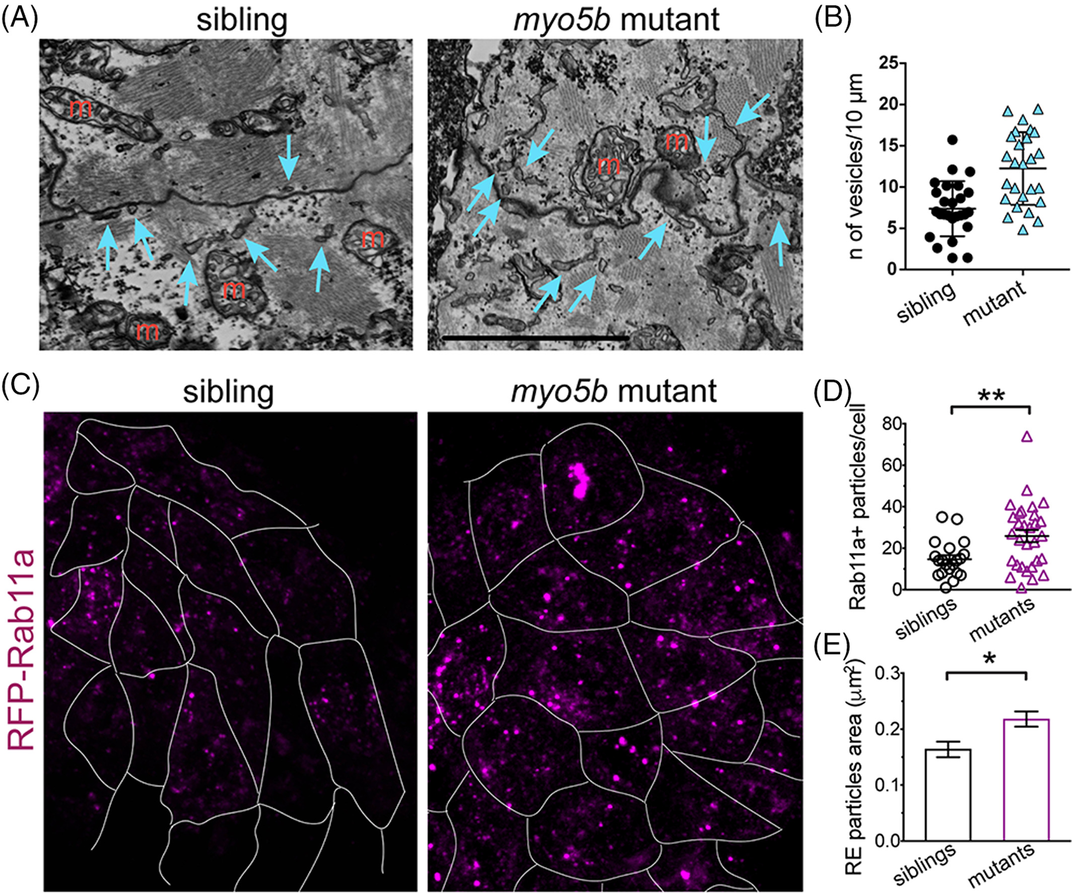
|
Fig. 3 myo5b mutant cardiomyocytes have increased recycling endosomes. A: Electron microscopic images of 36 hpf sibling and uq23ks mutant cardiomyocytes showing nonmitochondrial vesicles and tubular structures (blue arrows). M, mitochondria; Scale bar = 2.5 μm. B: Graph quantifying vesicles and tubular structures in siblings vs. mutant cardiomyocytes within 1 μm of the plasma membrane; mean ± SEM. C: Confocal imaging of recycling endosomes (labeled with RFP‐Rab11a) in sibling and mutant cardiomyocytes at 48 hpf. Cardiomyocyte membranes demarcated with white outline. D: Dot plot of the number of recycling endosomes per cell (as determined by RFP‐Rab11a‐postive particles), showing significantly more recycling endosomes in cardiomyocytes of uq23ks mutants vs. siblings. E: Histograms of recycling endosome size, showing significantly larger endosome size, consistent with endosome fusion.