
|
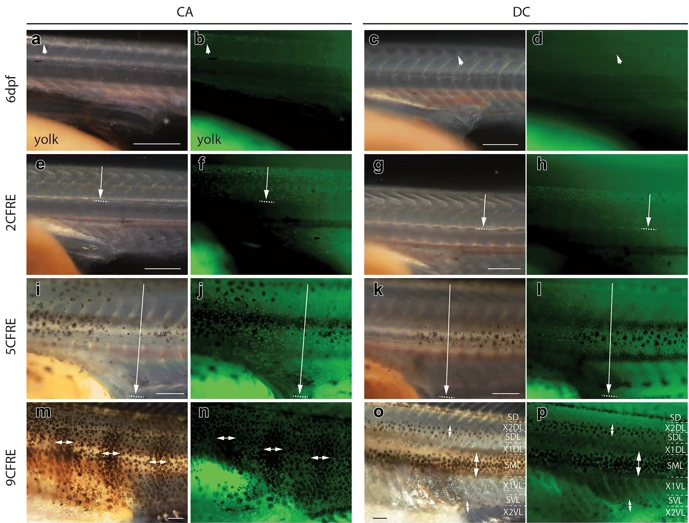
Fig. 4
Xanthophore distributions in C. azureus and D. compressiceps larvae. a–d Xanthophores are restricted to the dorsal midline in C. azureus and D. compressiceps at 6dpf (arrows) as visualized with white light (a, c) or 488 nm fluorescent light (b, d). e–h Xanthophores extend from dorsal midline to horizontal myoseptum (arrow) in C. azureus and D. compressiceps as visualized with white light (e, g) or 488 nm fluorescent light (f, h). i–l Xanthophores extend from dorsal to ventral midlines (arrow) in C. azureusand D. compressiceps as visualized with white light (i, l) or 488 nm fluorescent light (j, l). m–nXanthophore density is greater under melanophore patches (double-headed arrows) in C. azureus at 17dpf/9CFRE. o–p Xanthophores are largely restricted to dark stripes (double-headed arrows) in D. compressiceps at 17dpf/9CFRE. Scale = 500 µm