
Fig. 3
- ID
- ZDB-IMAGE-190916-2
- Publication
- Toms et al., 2019 - Phagosomal and mitochondrial alterations in RPE may contribute to KCNJ13 retinopathy
- All Figures
- Figures for Toms et al., 2019
|
Fig. 3
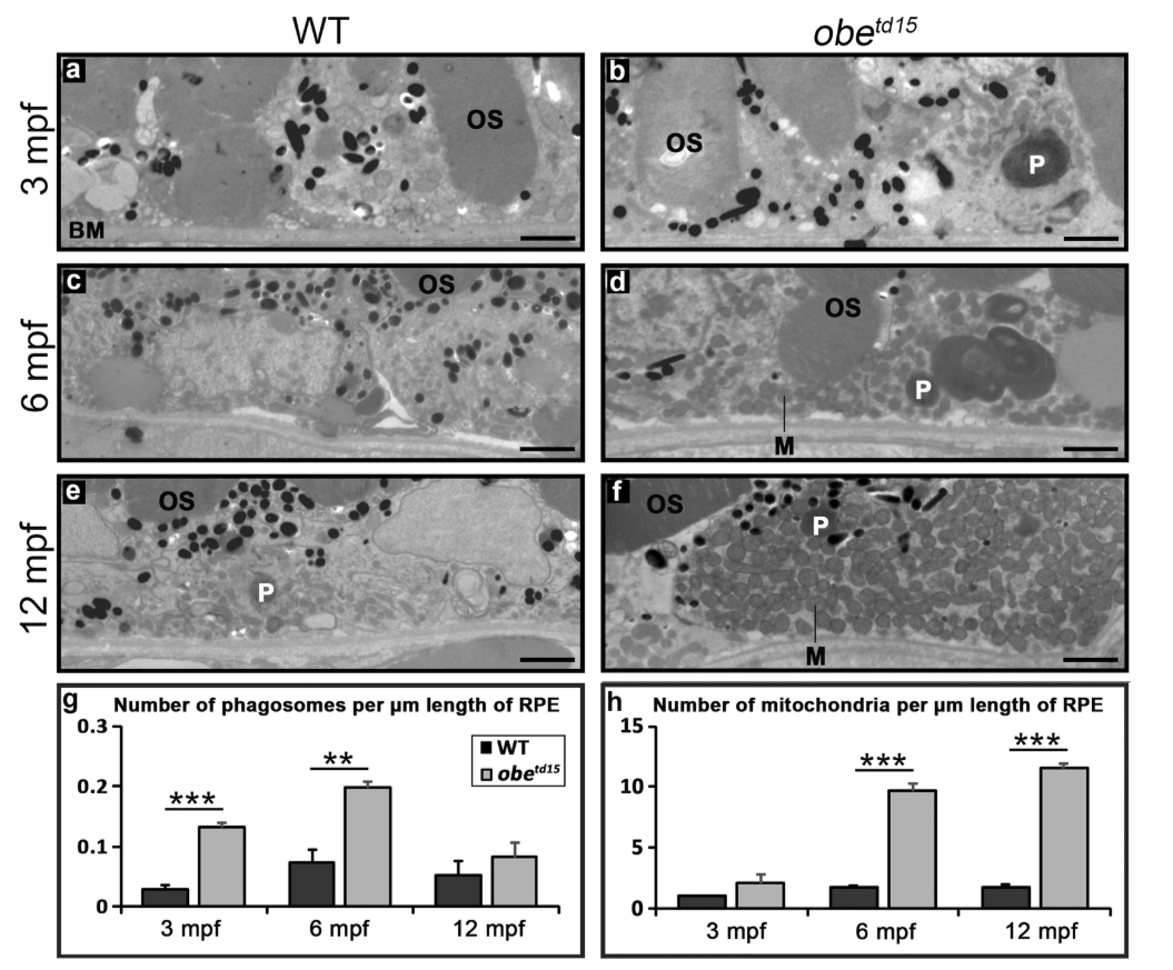
Increased mitochondrial abundance and altered phagosome clearance in the retinal pigment epithelium (RPE) of obetd15zebrafish. TEM cross-sections of the RPE show little difference at 3 months post fertilization (mpf) (a,b), while at 6 mpf (c,d) and 12 mpf (e,f) there is a significant increase in the number of mitochondria (M) in obetd15 compared to wild-type (WT) eyes. Additionally, there is a significant increase in the number of phagosomes (P) in obetd15 eyes at 3 mpf and 6 mpf. Bar charts displaying numbers of phagosomes and mitochondria per µm length of RPE are shown in (g,h). OS, photoreceptor outer segments; BM, Bruch’s membrane. Results are mean ± SEM (n = 3). **p < 0.01, ***p < 0.001. Scale bars = 2 µm.