
|
Fig. 3
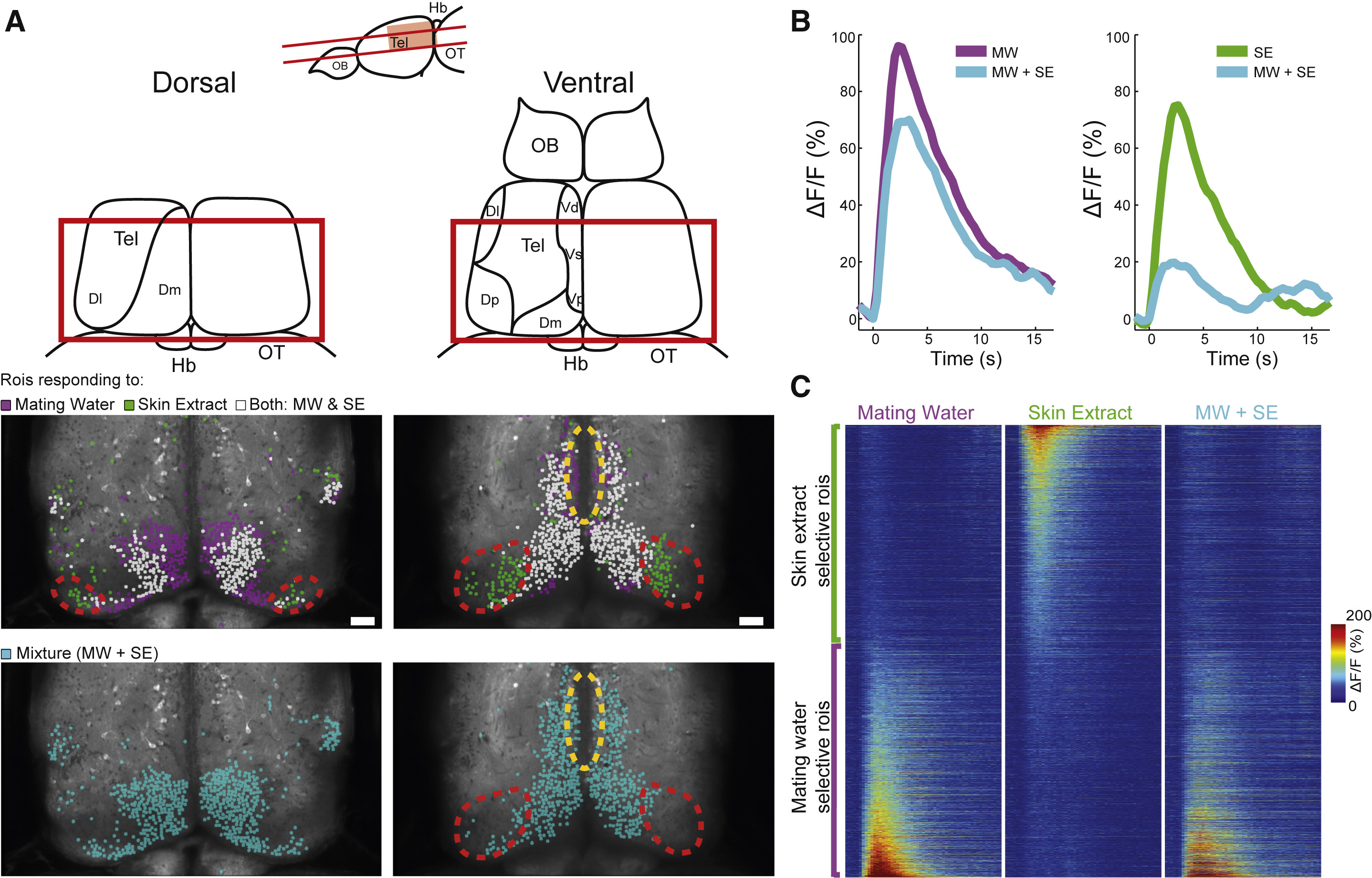
Skin Extract and Mating Water Elicit Responses in Both Specific and Overlapping Telencephalic Areas
(A) Anatomical reference (upper panel schematic) of 2-photon imaged brain regions (lower panel images) in 34-day-old juvenile tuba1a:GCaMP6s zebrafish. Each colored dot represents an roi activated in response either to one stimulus specifically (magenta, mating water; green, skin extract), to both stimuli (white), or to the mating-water and skin-extract stimulus mixture (cyan). Red-dotted regions contain skin-extract-selective rois that were suppressed when given the stimulus mixture (Dp and posterior-lateral Dm). Yellow-dotted region delineates Vs, which contain mating-water-selective rois, whose activity persisted when given the stimulus mixture.
(B) Left panel: mean ΔF/F signal of mating-water-selective rois in the telencephalon (n = 4,104 rois, n = 6 fish). Right panel: mean ΔF/F signal of skin-extract-selective rois in the telencephalon (n = 4,109 rois, n = 6 fish).
(C) ΔF/F signal of skin-extract- (top rows) or mating-water- (bottom rows) selective rois in a 20-s window post-delivery of mating water, skin extract, or mixture stimulus (n = 8,213 rois, n = 6 fish).
See also Figure S3. Tel, Telencephalon; OB, olfactory bulb; Hb, habenula; OT, optic tectum; DI, lateral zone of the dorsal telencephalic area; Dm, medial zone of the dorsal telencephalic area; Dp, posterior part of the dorsal telencephalic area; Vs, supracommisural nucleus of the ventral telencephalic area; and Vp, postcommisural nucleus of the ventral telencephalic area. Scale bar, 50 μm.