
|
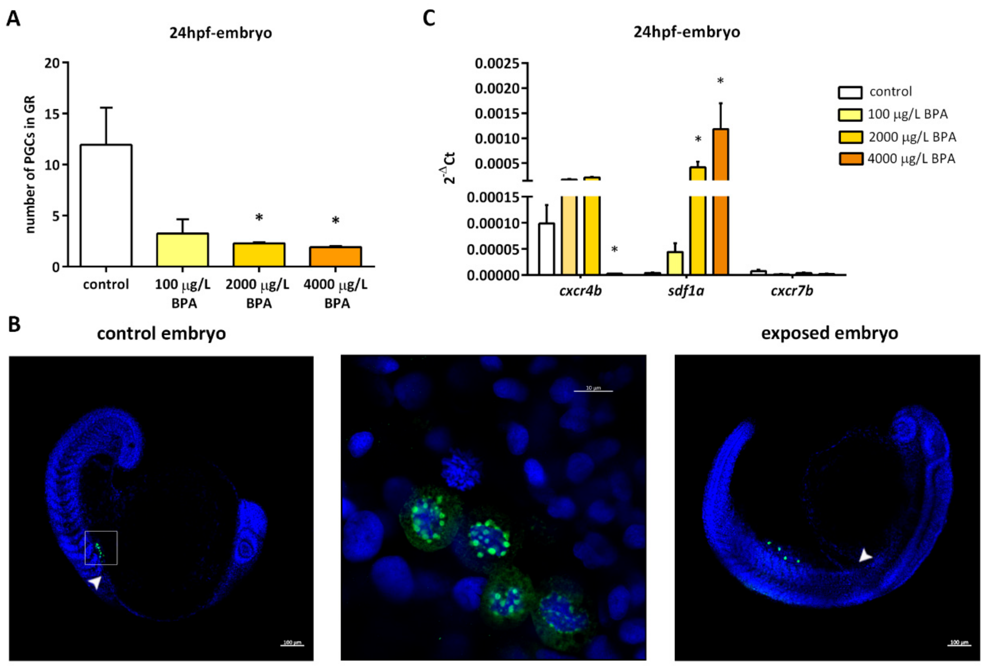
Fig. 1
(A) Number of primordial germ cells (PGCs) observed in the genital ridge of embryos at 24 h post fertilization (hpf). Bars represent mean of PGCs in 5 embryos at 24 hpf in 3 different experiments (n = 3). Asterisks indicate significant differences (p < 0.05) when compared to control groups. (B) Location of PGCs (green fluorescence) by confocal imaging in 24 hpf embryos. In control embryos (left picture), they were located within the genital ridge (arrowhead); scale bar 100 µm. The inset region is enlarged in the middle picture, in which germ cells are marked with Vasa (green spots surrounding nuclear area stained with DAPI); scale bar 10 µm. In 24 hpf embryos exposed to 4000 µg/L BPA (right picture), PGCs appeared outside the genital ridge (arrowhead); scale bar 100 µm. (C) Relative expression of genes involved in primordial germ cell migration in the genital ridges of control and BPA-exposed embryos. Expression levels relative to 18 S rRNA were calculated using 2−ΔCt method in three independent experiments (n = 3). Asterisks indicate significant differences (p < 0.05) when compared to control embryos.