
|
Fig. 1
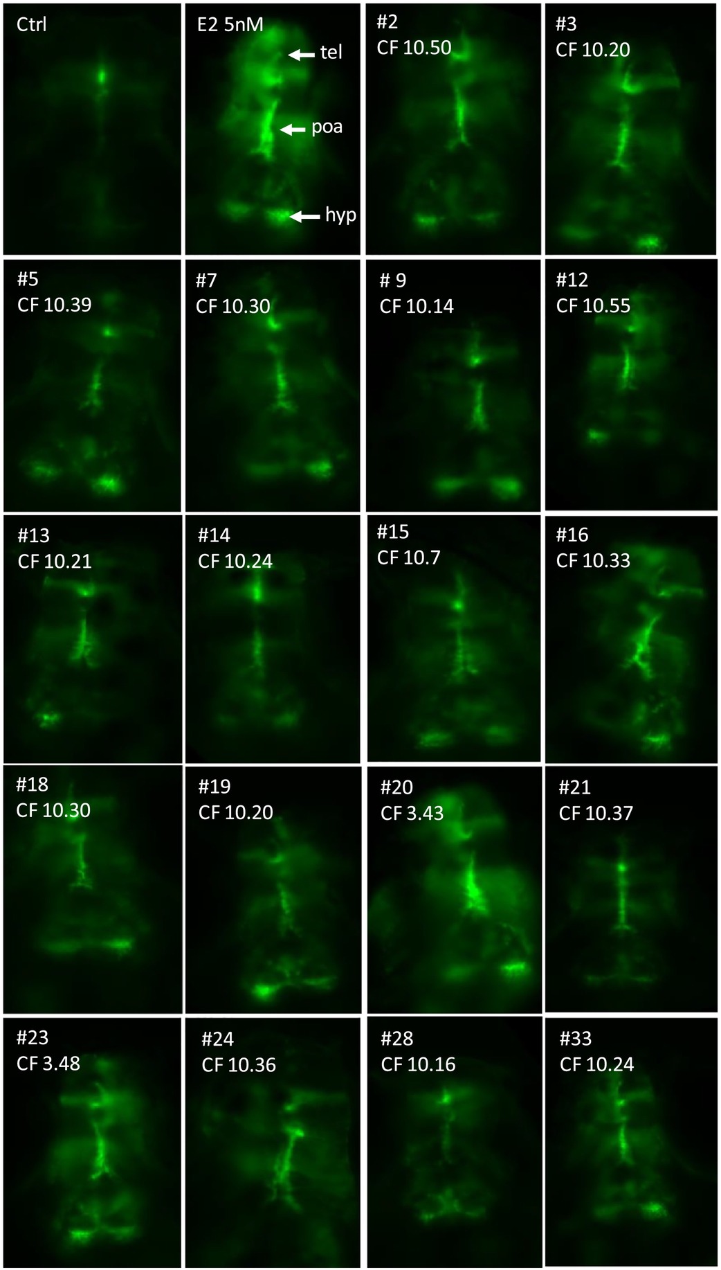
In vivo imaging of the brain of 4-dpf old live transgenic cyp19a1b-GFP zebrafish embryos exposed to waste water (WW) and surface water (SW) sample extracts that were all able to induce a significant response above control in the EASZY assay. Fluorescent signal (green color) reveals induced GFP expression in the developing brain. Dorsal views (anterior to the top) of the telencephalon (tel), preoptic area (poa), and the caudal hypothalamus (hyp). For each water samples, the concentration factor (CF) of tested extract is indicated. CTRL: solvent control, E2 = 17β-estradiol, # = sample number. Scale bar = 100 μM. (For interpretation of the references to color in this figure legend, the reader is referred to the web version of this article.)