
Image
|
Figure Caption
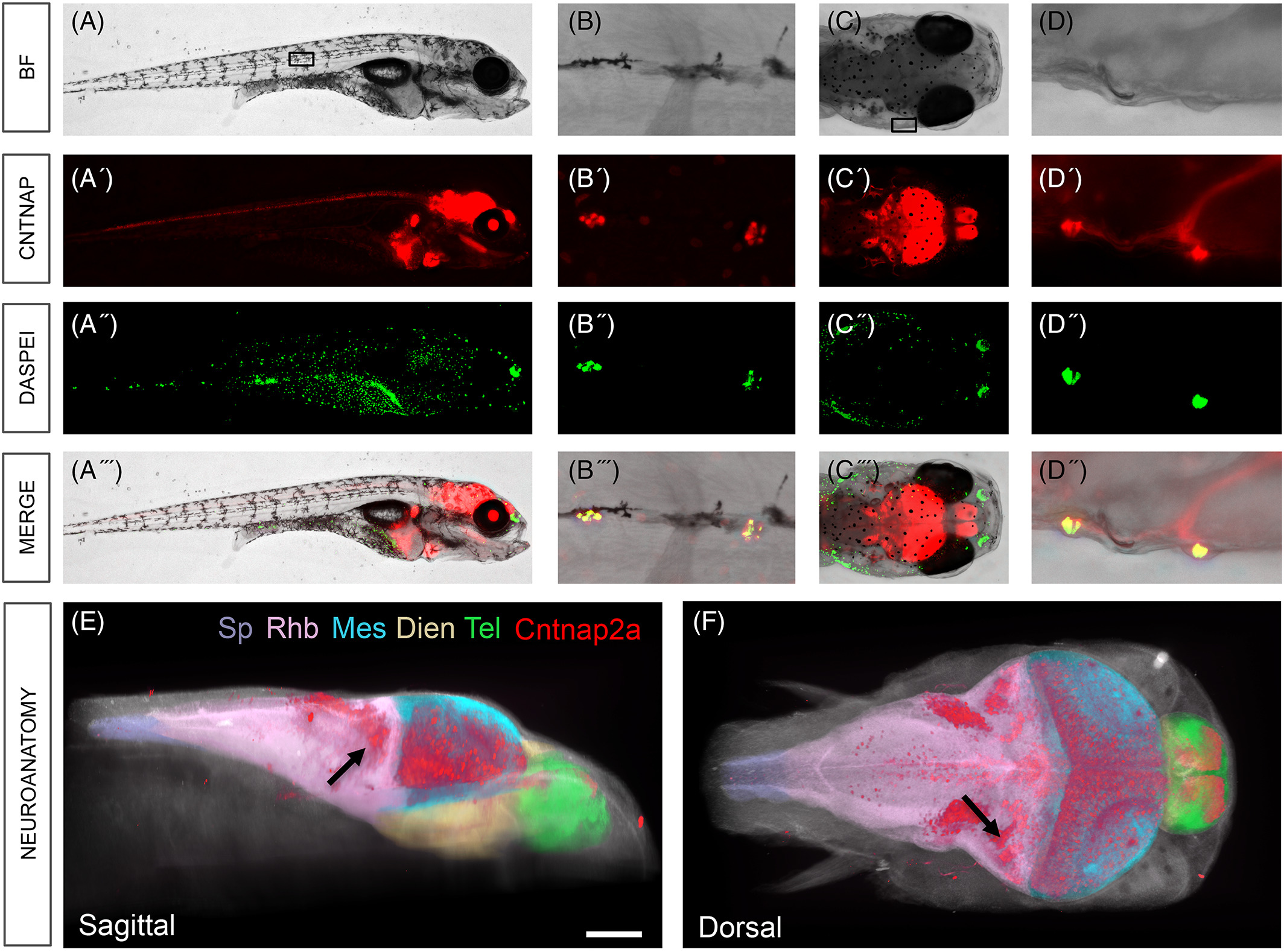
Fig. 4
Cntnap2a colocalizes with DASPEI neuromast stain. Whole body view (A‐A′′′) and zoom (B‐B′′′) show that Cntnap2a‐mCherry (red) and neuromasts of lateral line (DASPEI, green) colocalize (merge, B′′′). Dorsal head view (C‐C′′′) and zoom (D‐D′′′) demonstrates colocalization of neuromasts and Cntnap2a labeled neurons that innervate the brain (merge, D′′′). E, Sagittal aspect. F, Dorsal aspect (SB = 100 μm). Spine (Sp, purple), rhombencephalon (Rbh, pink), mesencephalon (Mes, blue), diencephalon (Dien, yellow), telencephalon (Tel, green), Cntnap2a:mCherry (red). E, Arrow, medial octavolateralis nucleus
Acknowledgments
This image is the copyrighted work of the attributed author or publisher, and
ZFIN has permission only to display this image to its users.
Additional permissions should be obtained from the applicable author or publisher of the image.
Full text @ Dev. Dyn.