
|
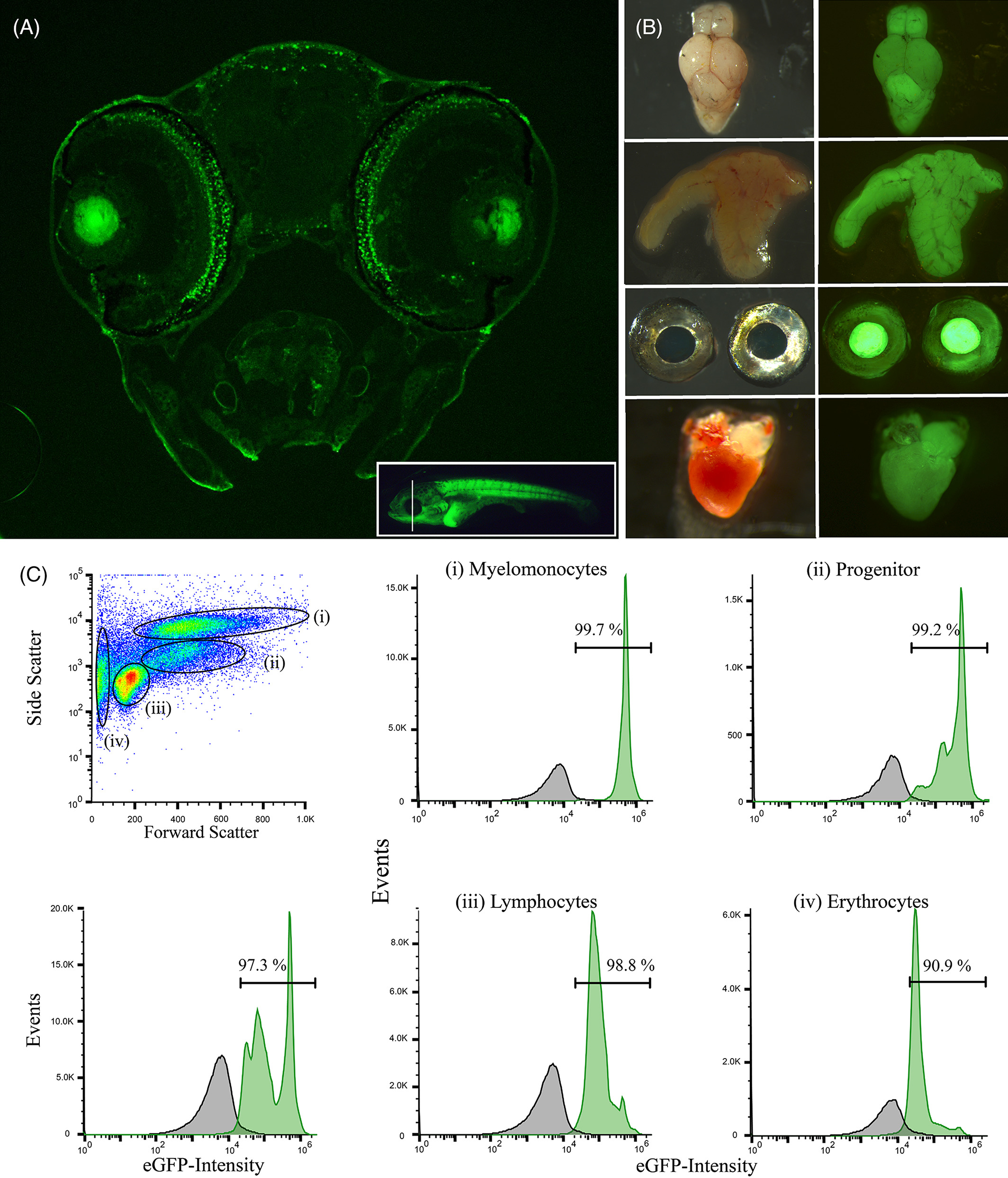
Fig. 2
Ubiquitin promoter driven transgene expresses GFP in different tissues. A, transverse section of a 14‐dpf‐old Ubi‐GFP transgenic surface fish. Inset picture indicates the location of the section. B, GFP expression in different organs of adult transgenic fish, from top to bottom: brain, liver, eyes, heart. C, Fluorescence activated cell sorting of head kidney cells from Ubi‐GFP transgenic A. mexicanus. Four distinct hematopoietic cell populations can be identified by forward scatter and side scatter: (i) myelomonocytes, (ii) progenitors, (iii) lymphocytes, and (iv) erythrocytes. Green indicates GFP signal from Ubi‐GFP transgenes, while dark grey indicates GFP signal from wild‐type control