
|
Fig. 3
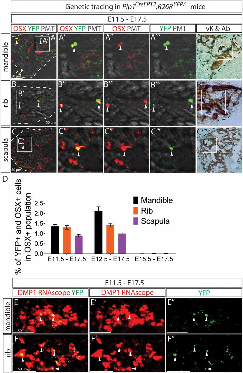
SCPs generate osteoprogenitor cells and osteocytes in facial region and trunk during murine embryonic development. (A–C) SCPs progeny in Plp1CreERT2;R26RYFP/+ embryos traced from E11.5 to E17.5 were positive for osteoprogenitor marker OSX in the ossified parts of mandible (A), rib (B), and scapula (C). The white arrowheads indicate double-positive cells. (D) Quantification of YFP+ cells among the OSX+ population. Data represent mean ± SEM where at least 3 embryos from independent litters were analyzed. (E and F) The same traced embryos were positive for Dmp1 RNA probe. A′′′′, B′′′′, and C′′′′ depict the same tissue sections as A–C but stained with von Kossa and Alcian blue (vK and Ab). The white dashed lines outline the mineralized portion of the bone.