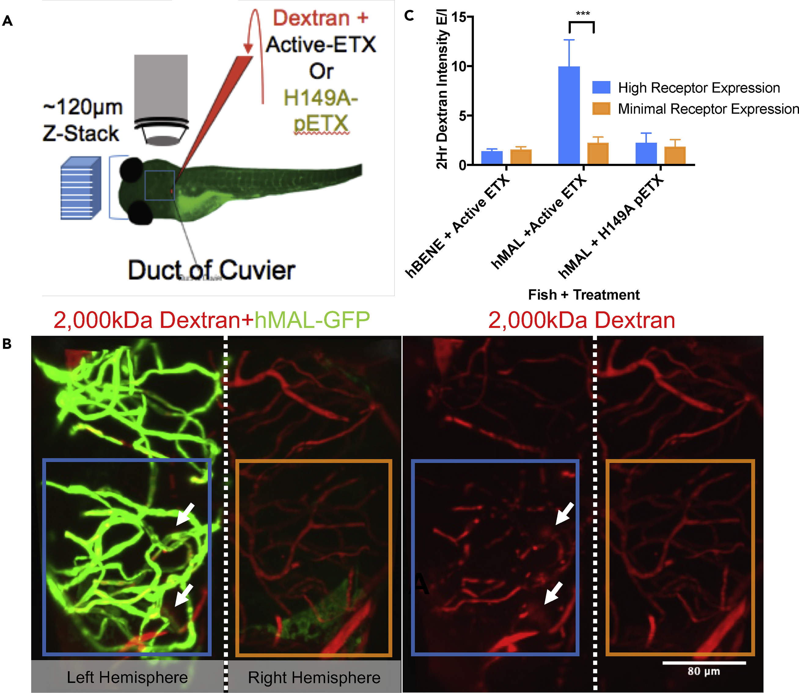
Fig. 4
Within Individual hMAL-GFP-Expressing Fish, Dextran Leaks Markedly More in Volumes of the Brain Populated by Receptor-Expressing Vessels than in Areas Without Receptor Expression
(A) Schematic of injection and imaging strategy.
(B) Confocal z stack of a representative fish transiently expressing hMAL-GFP (green) injected with active toxin and 2,000-kDa rhodaminedextran (red) and an adjacent image of just the red dextran channel. Because of the chimeric nature of expression most fish only expressed receptor in discrete parts of the brain, and as depicted, often in half of the brain. White arrows indicate regions of evident leakage. Blue and orange boxes represent the brain quadrants quantified in (C).
(C) Quantification of E/I ratio at 2 HPI between volumes of the brain that have high levels of dextran expression (blue) and those with little dextran expression (orange) for each fish in each condition: hBENE transients injected with active ETX (n = 5/side of brain), hMAL transients injected with active ETX (n = 6/side of brain), hMAL transients injected with H149A mutant pETX (n = 4/side of brain). Significant difference between these two sides of the brain are only seen in the hMAL + active ETX condition (two-way ANOVA with Tukey's test, p = 0.0008). Data are represented as mean ± SEM.

