Fig. 3
Co-injection of Activated ETX and 2,000-kDa RhodamineDextran Reveals Pronounced BBB Leakage in hMAL-Expressing Fish
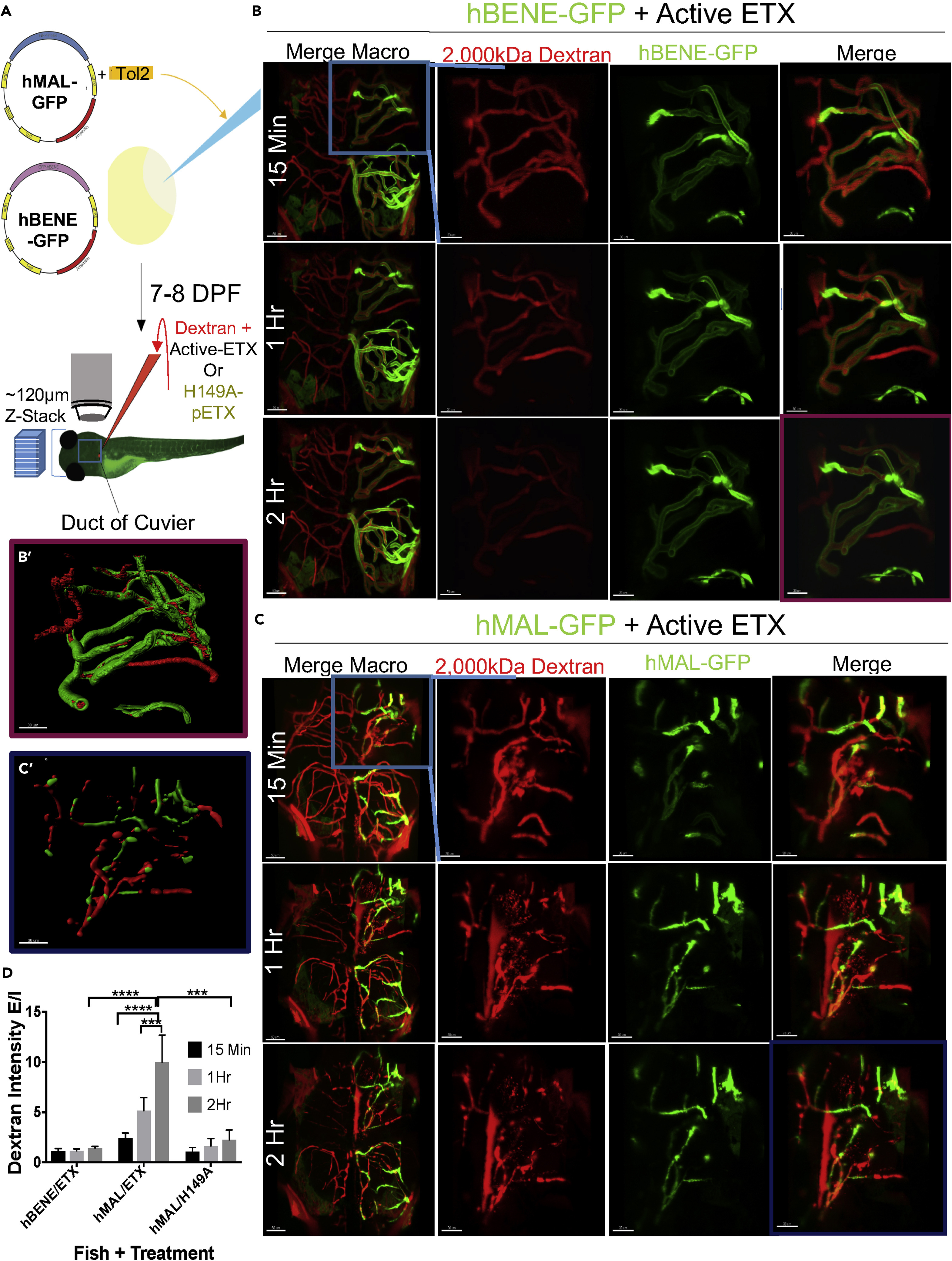
(A) Schematic depicting experimental procedure.
(B) The 40× confocal z stacks of the vasculature in the anterior dorsal quadrant of the brain in a representative hBENE-GFP transiently expressing control zebrafish. hBENE-GFP receptor is in green, the 2,000-kDa rhodamine dextran is in red, “merge” images show both channels, and “Merge Macro” images show full-brain neurovasculature. The blue box in “Merge Macro” highlights the area focused in for the other images; 15 min, 1 h, and 2 h indicate the time z stack imaging commenced after toxin or dextran injection. Scale bar, 50 μm in “Merge Macro” and 30 μm in the rest of the images.
(C) Same as (B) but in an hMAL-GFP-expressing fish. (B′) Imaris “surface” rendering of hBENE-GFP in green and rhodamine dextran in red used for quantification of GFP sum intensity and total “intravascular” rhodamine sum intensity within the lumen of blood vessels at 2 h post injection. (C′) Same as (B′) but in hMAL-GFP fish.
(D) Quantification of dextran leakage ascertained by determining extravascular dextran sum intensity divided by intravascular sum intensity (“dextran E/I”) at 15 min, 1 h, and 2 h post injection. Data included from experiment using hBENE-GFP fish injected with active ETX (n = 5), hMal-GFP fish injected with active ETX (n = 6), and hMal-GFP fish injected with mutant control H149A pETX (n = 4).
Data are represented as mean ± SEM. Statistical analysis: two-way ANOVA with Tukey’s test (***p < 0.001, ****p < 0.0001).

