
|
Fig. 4
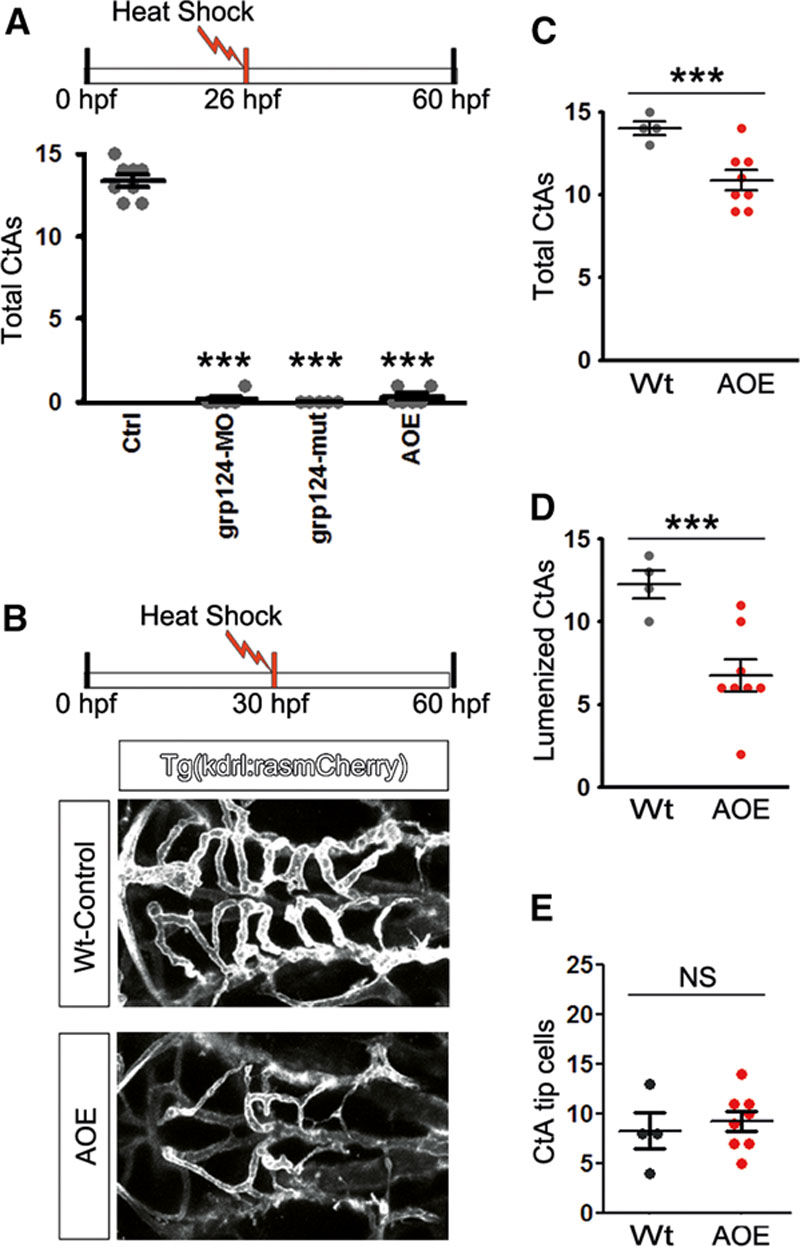
Brain vascular maturation and stability are compromised in zebrafish upon Axin1iEC−OE. A, Top: Experimental design. Bottom: Quantification of the number of central arteries (CtAs) formed at 60 h postfertilization (hpf) in Tg(kdrl:rasmCherry; Wt, Ctrl), Tg(adgra2s984;kdrl:rasmCherry; gpr124-mut), grp124-morpholino-injected Tg(kdrl:rasmCherry; grp124-MO) at the 1-cell stage or Tg(hsp70l:Mmu.Axin1-YFP;kdrl:rasmCherry) zebrafish embryos following heat-shock induced Axin1 overexpression at 26 hpf (AOE). ***P<0.001. B, Confocal micrographs of the central arteries in Wt or AOE zebrafish embryos at 60 hpf, exposed to heat-shock–induced AOE at 30 hpf. C–E, Quantification of the total number of CtAs (C), lumenized CtAs (D), or CtA tip cells (E) from the experiment shown in B. n=4–8 as indicated. NS indicates nonsignificant. ***P<0.001.