
Fig. S7
- ID
- ZDB-IMAGE-190816-7
- Publication
- Parker et al., 2019 - A Hox-TALE regulatory circuit for neural crest patterning is conserved across vertebrates
- All Figures
- Figures for Parker et al., 2019
|
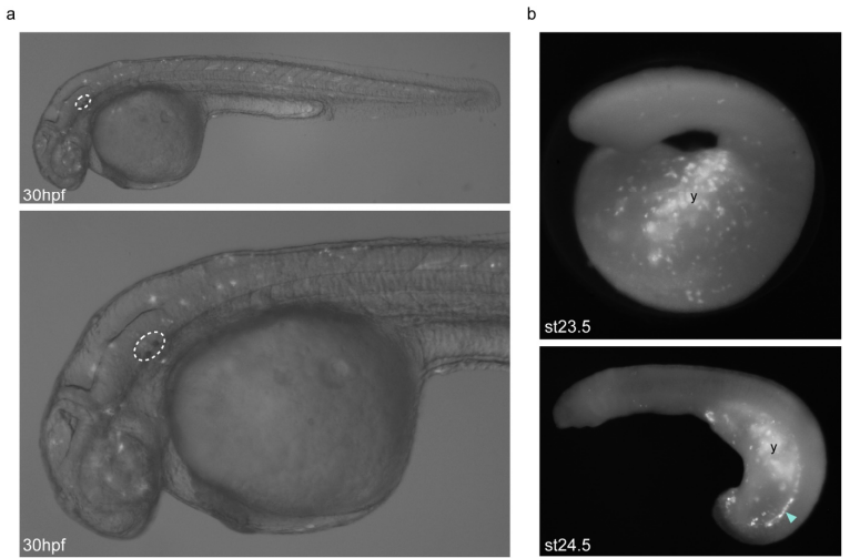
Fig. S7
Background GFP expression driven by the empty HLC vector in zebrafish and lamprey embryos. a, Lateral views of 30hpf/prim-16 stage transient transgenic zebrafish embryos injected with the empty HLC vector using Tol2-mediated transgenesis, showing low-intensity mosaic GFP expression in multiple tissue types including neurons and muscle cells. The otic vesicle is circled. b, Lateral views of st23.5 and st24.5 transient transgenic lamprey embryos injected with the empty HLC vector using I-SceI-mediated transgenesis, showing mosaic GFP expression in the yolk (y) and ectoderm, as well as cells lying dorsal to the yolk (arrowhead). GFP-expressing embryos shown are representative of the expression potential of the reporter construct in each case, as inferred from screening many (typically more than 100) injected embryos.